What is histopathology?
Histopathology (from histos, tissue, and pathos, disease) is the examination of tissue under a microscope to diagnose disease. A clinician removes a sample of tissue — a small needle-core biopsy, an endoscopic pinch, or a whole surgical resection — and sends it to the histopathology laboratory, where it is processed into thin, stained sections on glass slides. A pathologist then examines those sections and issues a report that guides treatment.
Histopathology differs from cytopathology, which looks at individual cells (for example, a Pap smear), in that it preserves the architecture of the tissue — how cells are arranged relative to one another. That architecture is often what distinguishes benign from malignant, or one tumour type from another. The histopathology report is frequently the definitive diagnosis on which surgery, chemotherapy or radiotherapy decisions are based.
Why histopathology matters
For most solid cancers, the histopathology report is the reference standard for diagnosis, tumour type, grade and, after surgery, the adequacy of excision margins. It also diagnoses inflammatory, infective and degenerative conditions across virtually every organ system. Because treatment decisions hang on it, the report must be both accurate and timely.
This is why laboratories track turnaround time (the interval from specimen receipt to authorised report) and quality as their core performance metrics. A delayed or inconsistent histopathology service holds up the whole clinical pathway; a fast, reliable one lets surgeons and oncologists act with confidence.
The histopathology workflow, end to end
The laboratory converts raw tissue into a readable slide through a fixed sequence of steps, each dependent on the one before:
- Specimen receipt & accessioning — logging and uniquely identifying every specimen.
- Grossing — describing, measuring and dissecting the specimen and sampling representative pieces into cassettes.
- Fixation — preserving the tissue, usually in neutral buffered formalin.
- Tissue processing — dehydration, clearing and paraffin infiltration.
- Embedding — orienting the tissue in a paraffin block.
- Microtomy — cutting thin sections, typically 3–5 µm.
- Floatation, mounting & drying — transferring sections onto slides.
- Staining — most commonly haematoxylin and eosin (H&E).
- Microscopy & reporting — examination and the issued report.
Because the steps are sequential, the whole report is only as fast and as good as its slowest or weakest stage. Most quality and turnaround problems trace back to a specific point in this chain.
Specimen receipt, grossing and fixation
Everything begins with correct identification. Each specimen is accessioned against the request form, and its identity must be controlled from this first moment to the final slide. At the grossing station the specimen is described, measured and dissected, and representative pieces are placed into labelled cassettes. Accurate sampling here determines what the pathologist ultimately sees, so good lighting, ergonomics and documentation matter.
Grossing also handles large volumes of formalin, a respiratory irritant and recognised hazard, so a grossing station with effective downdraft or backdraft fume extraction is a genuine safety requirement, not a luxury. Fixation itself — typically immersion in 10% neutral buffered formalin — stabilises proteins and stops autolysis. Adequate fixation time is essential; tissue that enters processing under-fixed processes poorly and forces repeats, so fixation is one of the most important and most overlooked controls in the whole workflow.
Tissue processing
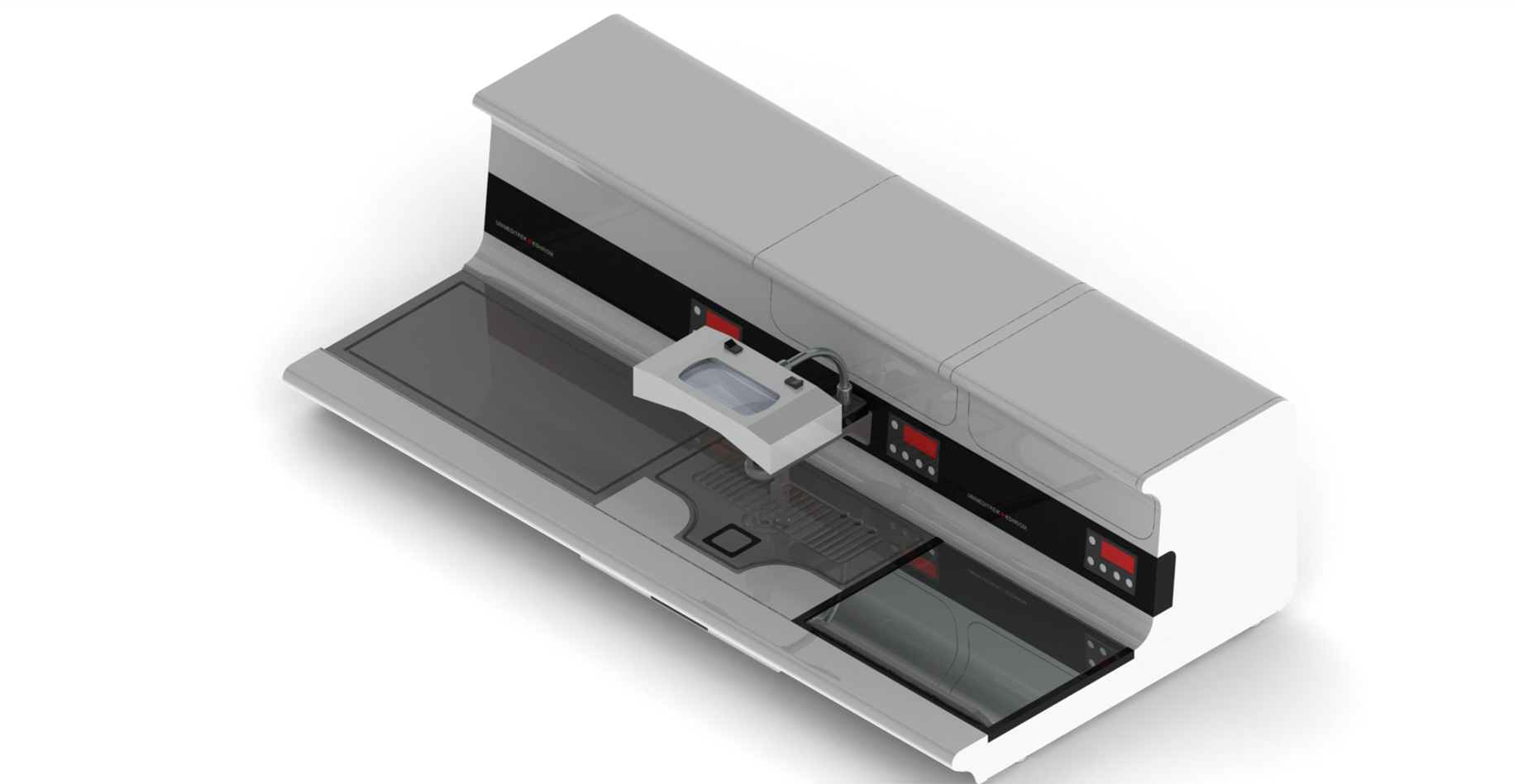
Fixed tissue is mostly water, and water and molten paraffin do not mix — so before a block can be made, the water must be replaced by wax. A tissue processor automates this through a programmed sequence: graded alcohols remove water (dehydration), a clearing agent such as xylene replaces the alcohol (clearing), and molten paraffin then infiltrates the tissue. Better processors apply vacuum and pressure to drive complete infiltration of dense, fatty or decalcified tissue.
Processing is the foundation of a sectionable block: only well-processed tissue cuts cleanly at 3–5 µm. Programmable, repeatable schedules — short and gentle for small biopsies, longer for large or fatty resections — make every run consistent, while reagent-management features extend reagent life safely and reduce both cost and fume exposure. See the Complete Guide to Tissue Processors for a deeper treatment.
Embedding
After processing, each piece of tissue is embedded in paraffin at an embedding station: it is placed in a mould, correctly oriented, and surrounded by molten wax over a cold plate that sets the block. Orientation is critical — a tubular structure embedded on its side, or a skin biopsy embedded flat, will not yield a representative section no matter how good the downstream steps are.
A good embedding station provides a heated paraffin reservoir, a warm working surface, a cold spot for rapid setting and a heated forceps well, laid out for continuous, comfortable work. Embedding quality directly governs how much diagnostic tissue the pathologist actually sees. See the Complete Guide to Tissue Embedding Stations.
Microtomy: section cutting
Microtomy is the art of shaving ribbons of tissue from the block at a uniform thickness, routinely 3–5 µm. The block is advanced against a sharp blade by a precise feed mechanism; motorised microtomes give a smooth, repeatable advance that reduces chatter and operator fatigue. Most faults blamed on "the instrument" are in fact blade or block faults — a blunt blade, an incorrect clearance angle, or a block that was not properly infiltrated or cooled.
The thin ribbons produced here are what carry the diagnostic information forward; uneven, thick or folded sections stain unevenly and can obscure morphology, so consistency at microtomy protects everything downstream.
Floatation, mounting and drying
Ribbons from the microtome are floated on warm water in a tissue floatation bath to relax folds and wrinkles, then picked up onto a glass slide. The water is held at a precise temperature just below the paraffin melting point — too hot over-expands and disrupts the section, too cool leaves folds — so a stable, even bath temperature has a direct effect on how clean the final slide looks.
Mounted slides are then dried on a slide warming table to drain residual water and bond the section to the glass before staining. Even, controlled heat prevents the drying artefacts and section loss that come from hot spots or excessive temperature. These two unglamorous steps quietly determine a large share of final slide quality.
Staining: H&E and special stains
Paraffin sections are almost colourless, so they must be stained to reveal structure. The routine workhorse is haematoxylin and eosin (H&E): haematoxylin stains cell nuclei blue-purple, eosin stains cytoplasm and connective tissue pink. The pathologist reads morphology through this stain, so consistency — the same colour and contrast on every slide, every shift — is the baseline for reliable diagnosis.
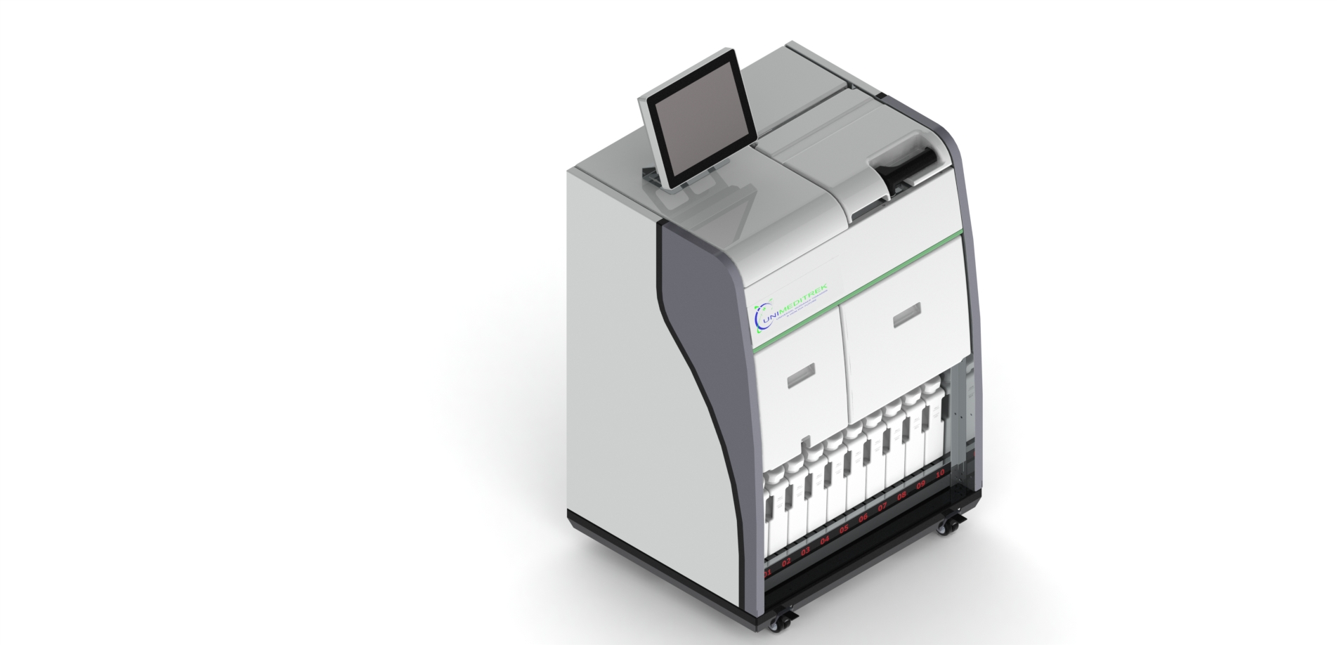
Beyond H&E, special stains highlight specific targets (for example, stains for connective tissue, micro-organisms or pigments), and immunohistochemistry uses antibodies to identify particular proteins for cancer typing. An automatic slide staining machine applies identical timing and sequence to every slide, removing the operator-to-operator variability of manual staining and freeing technologists. See the Complete Guide to Slide Staining Machines.
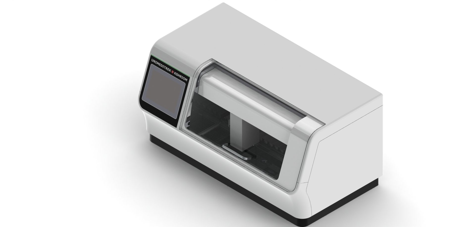
Frozen section and the cryostat
Sometimes a surgeon needs an answer during an operation — for example, whether a tumour margin is clear. Frozen section delivers a provisional diagnosis within minutes: fresh, unfixed tissue is frozen in a refrigerated cryostat microtome, sectioned, rapidly stained and read while the patient is still in theatre. It is one of the most time-critical tasks in pathology, demanding stable chamber temperature, a sharp blade and a disciplined workflow. See the Complete Guide to Cryostat Microtomes.
Microscopy, reporting and digital pathology
The finished slide is examined under a microscope, and the pathologist integrates the findings into a report: the diagnosis, tumour type and grade where relevant, and margin status after resection. Increasingly, slides are also being digitised — scanned into high-resolution whole-slide images that can be viewed on a screen, shared for second opinions, archived, and analysed with image-analysis and AI tools.
Digital pathology does not change the physical workflow that produces the slide — fixation, processing, microtomy and staining still have to be excellent — but it is changing how slides are reviewed, taught and quality-assured, and it is one of the most important trends shaping the field.
Equipment in a histopathology laboratory
A complete histopathology laboratory is built around the workflow above. The core equipment set is: a grossing station, a tissue processor, a tissue embedding station, a microtome, a tissue floatation bath, a slide warming table and an automatic slide staining machine — plus a cryostat microtome wherever frozen sections are performed.
Selecting and sizing this equipment to the expected case load, and planning the laboratory layout and utilities around a one-way workflow, is itself a discipline — covered in the Complete Guide to Histopathology Laboratory Design.
Quality, turnaround and the future
A good histopathology service is judged on accuracy and turnaround time, both of which depend on disciplined fixation, reproducible processing and staining, well-maintained equipment, and a smooth flow with minimal rework. Accreditation frameworks such as NABL formalise this with documented procedures, calibration, internal quality control and traceable records.
Looking ahead, the field is being reshaped by automation, digital and computational pathology, and a growing emphasis on standardisation. The fundamentals, however, do not change: every digital image and every AI result still rests on a well-fixed, well-processed, well-cut and well-stained section. Mastering the physical workflow remains the foundation of histopathology.
Frequently asked questions
What is the difference between histopathology and cytopathology?
Histopathology examines tissue sections and preserves tissue architecture (how cells are arranged), while cytopathology examines individual cells, such as in a Pap smear. Architecture is often what distinguishes benign from malignant.
What are the main steps in the histopathology workflow?
Specimen receipt and accessioning, grossing, fixation, tissue processing, embedding, microtomy, floatation and drying, staining, and finally microscopy and reporting.
At what thickness are histopathology sections cut?
Routine paraffin sections are typically cut at 3–5 µm, which is thin enough for light to pass through and for cellular detail to be seen after staining.
What is H&E staining?
Haematoxylin and eosin staining is the routine stain in histopathology: haematoxylin stains nuclei blue-purple and eosin stains cytoplasm and connective tissue pink, revealing tissue structure for diagnosis.