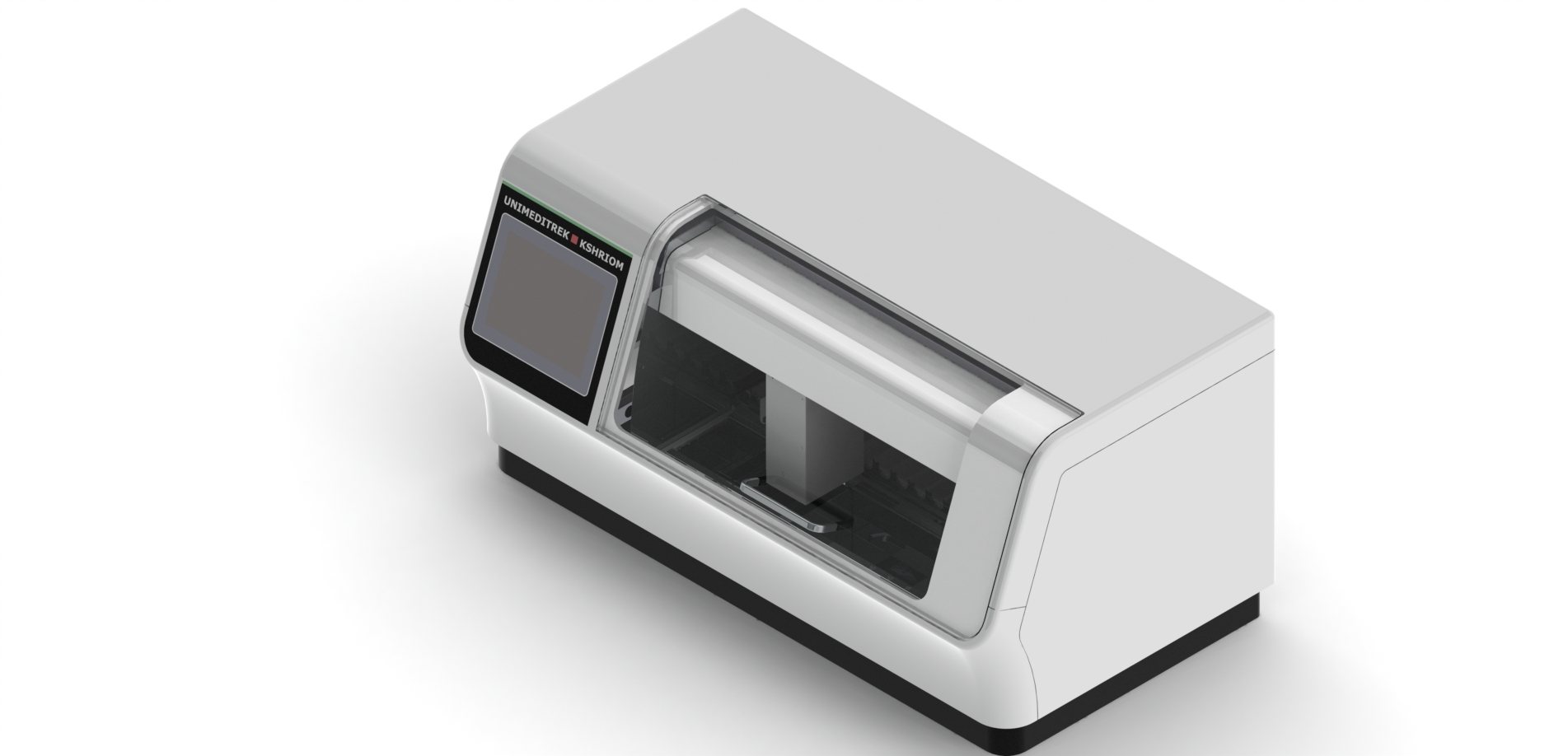
What a cryostat is and when it is used
A cryostat microtome is essentially a microtome housed inside a freezer cabinet. Its main job is frozen section — rapid, intra-operative diagnosis. Unlike the routine paraffin workflow, which takes hours, frozen section produces a readable slide in minutes by freezing the tissue solid instead of processing and embedding it in wax.
Typical uses include checking surgical margins, confirming that a sampled lesion is representative, and identifying tissue type to guide the next surgical step. It is one of the most time-critical tasks in pathology, and the cryostat is built around speed and stability.
How a cryostat works
Fresh tissue is mounted on a chuck with a freezing medium and frozen rapidly — a fast-freeze shelf or heat-extractor brings it to sectioning temperature quickly, which keeps ice crystals small and preserves morphology. The microtome inside the chamber then cuts thin sections, usually around 4–8 µm, which are picked up onto a slide, rapidly stained (commonly a rapid H&E or toluidine blue) and read immediately.
Because the whole instrument sits at sub-zero temperature, both the chamber and the specimen are temperature-controlled, and a sharp blade plus a stable temperature are what separate a clean diagnostic section from a torn, useless one.
Chamber temperature by tissue type
The right cutting temperature depends on tissue composition, and getting it wrong is the most common reason sections fail:
- Fatty / lipid-rich tissue (e.g. breast) cuts best colder, often around −25 °C or below, because fat stays soft.
- Lean, cellular tissue (e.g. lymph node) cuts at warmer settings, often around −15 to −20 °C.
Most cryostats let you set chamber and specimen-head temperatures, and experienced technologists adjust to the block in front of them. A cabinet that holds its set temperature stably — without large swings as the door opens during a busy list — is therefore a core requirement.
The intra-operative workflow and turnaround
Frozen section is a relay against the clock: the specimen arrives fresh from theatre, is oriented and frozen, sectioned, stained and read, and the result is phoned back to the surgeon — often within ten to twenty minutes. Anything that adds delay or forces a re-cut costs the patient time under anaesthesia, so a disciplined, rehearsed workflow and a reliable instrument matter enormously. This sits within the wider diagnostic pathway described in the Complete Guide to Histopathology.
Features that matter
- Stable, accurate chamber temperature with independent specimen-head cooling.
- Fast-freeze shelf / heat extractor to bring blocks to temperature quickly and limit ice-crystal artefact.
- Anti-roll plate and a precise feed for flat, smooth sections.
- Automatic defrost management that does not disrupt a case in progress.
- Decontamination features (e.g. UV) for biosafety between cases.
- Smooth, reliable microtome mechanism and easy blade change for uptime during a list.
Safety with fresh, unfixed tissue
Frozen section works on fresh, unfixed tissue, which may be infectious, so a cryostat is a biosafety device as well as a microtome. Good practice includes treating all specimens as potentially infectious, routine chamber decontamination, careful blade handling at low temperature, and avoiding frozen section on samples with a known high-risk infectious agent. These are not optional extras — they are part of running the instrument correctly.
Common frozen-section problems
- Holes / lacework (ice-crystal artefact) — tissue frozen too slowly; use the fast-freeze shelf or heat extractor.
- Sections curling or not flattening — anti-roll plate misaligned or wrong temperature for the tissue.
- Chatter or thick/thin sections — blunt blade, incorrect clearance angle, or block not at the right temperature.
- Tearing — tissue too cold for its type, or a damaged blade edge.
As with paraffin microtomy, most faults are blade, block-temperature or technique issues rather than the cabinet itself.
Frequently asked questions
What is a frozen section used for?
Frozen section gives a rapid, provisional diagnosis during surgery — most commonly to check whether a tumour margin is clear, to confirm a sampled lesion is representative, or to identify tissue type so the surgeon can decide the next step while the patient is still in theatre.
What temperature should a cryostat be set to?
It depends on the tissue. Fatty, lipid-rich tissue such as breast cuts best colder (around −25 °C or below), while lean cellular tissue such as lymph node cuts at warmer settings (around −15 to −20 °C). A stable chamber temperature matters as much as the exact value.
Why are there holes or a lacework pattern in my frozen section?
That is ice-crystal artefact, caused by freezing the tissue too slowly. Using the fast-freeze shelf or heat extractor to freeze rapidly keeps ice crystals small and preserves morphology.
Is a cryostat a biosafety hazard?
Yes — it sections fresh, unfixed tissue that may be infectious. Treat all specimens as potentially infectious, decontaminate the chamber routinely (UV where available), handle blades carefully, and avoid frozen section on samples with known high-risk infectious agents.