What slide staining does
Staining gives contrast to a transparent tissue section. The routine stain is haematoxylin and eosin (H&E): haematoxylin stains cell nuclei blue-purple and eosin stains cytoplasm and connective tissue shades of pink. The pathologist reads architecture and cellular detail through this colour and contrast, so the consistency of the stain — the same result on every slide, every shift — is the baseline for reliable diagnosis.
Beyond H&E, laboratories run special stains (for connective tissue, micro-organisms, pigments and so on) and immunohistochemistry (IHC), which uses antibodies to identify specific proteins for cancer typing. The principles of standardised, timed, reproducible application are the same across all of them.
Manual vs automatic staining
Manual staining moves slide racks by hand through a row of reagent dishes, relying on the technologist to control dip time and sequence. It is flexible and cheap to start, but it is operator-dependent: timing drifts between people and across a busy day, reagents carry over, and results vary.
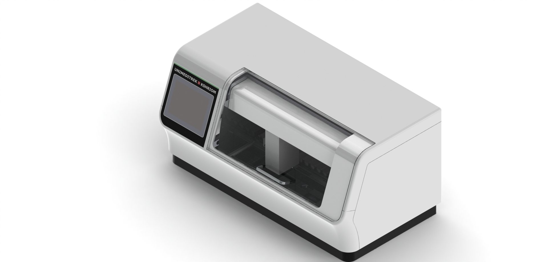
An automatic slide staining machine removes that variability. A programmed transport mechanism takes each rack through the exact same dip times and sequence, so slide 1 and slide 500 look identical. It also frees skilled staff from standing over reagent baths, controls reagent use, and contains solvent fumes. For any laboratory past a low daily volume, automation pays back in consistency, throughput and reproducibility.
Types of slide stainer
Stainers differ in how slides move through the reagents:
- Linear / continuous stainers feed racks through stations in a steady flow — well suited to high, steady H&E volumes.
- Batch / carousel stainers move a rack through a circular or programmed set of stations — flexible for mixed H&E and special-stain protocols.
- Robotic-arm stainers use a programmable arm to place racks into any station in any order, giving the most protocol flexibility.
Separately, stainers are categorised by purpose: routine H&E, special stains, and IHC stainers (often integrated with antigen retrieval). Many laboratories run a dedicated H&E stainer for volume plus a flexible platform for specials.
Features that matter
When comparing machines, weigh:
- Programmable protocols — multiple stored protocols with independent station times, so H&E and specials can run without re-plumbing.
- Reagent management — controlled volumes, station agitation and reminders that keep results consistent and reduce waste.
- Throughput — racks/hour matched to your real workload, not a headline figure.
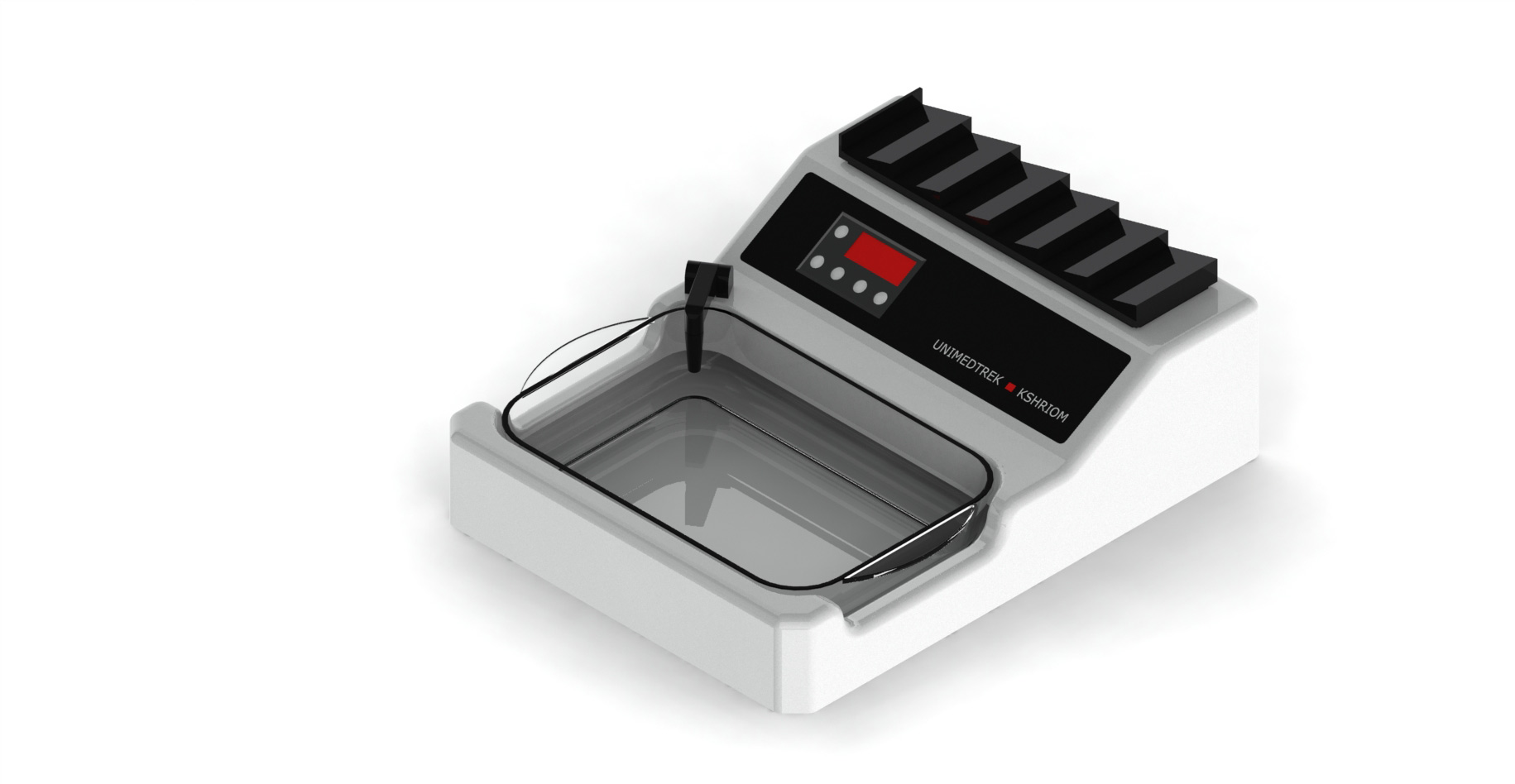
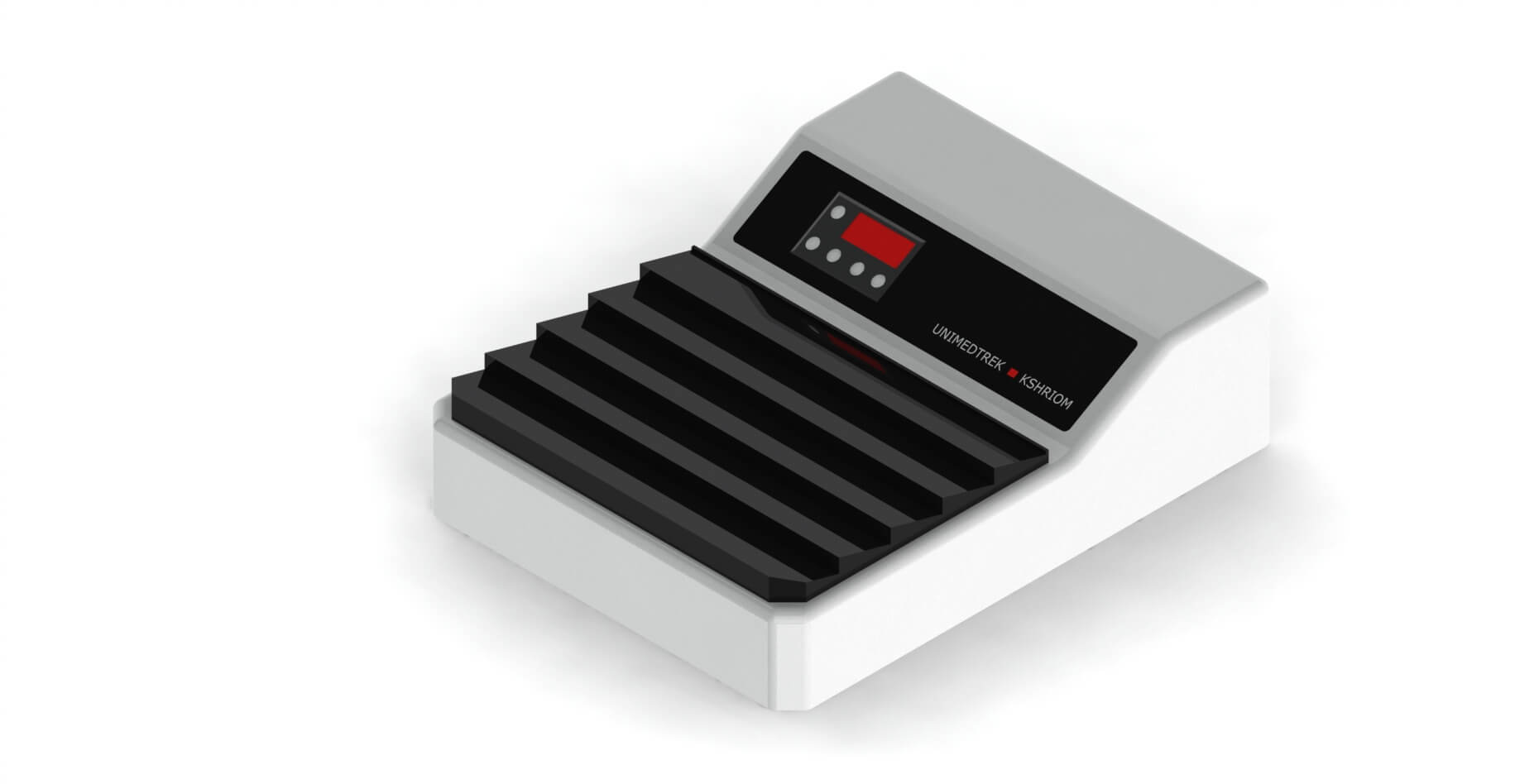
- Integrated drying / baking — on-board ovens that remove a manual step.
- Fume control — extraction or carbon filtration for xylene and alcohols.
- Walk-away operation — start-delay and continuous loading so staff are not tied to the bench.
Common staining problems and causes
Most staining faults trace to reagents, timing or upstream steps rather than the machine itself:
- Too pale or too dark — exhausted or fresh-but-mistimed haematoxylin/eosin, or incorrect dip times.
- Uneven or patchy staining — poor agitation, incomplete dewaxing, or sections not fully dried/adhered before staining.
- Contamination / floaters — carry-over between baths; scheduled reagent changes and bath cleaning fix it.
- Weak nuclear detail — over-differentiation, hard water, or under-fixed/over-processed tissue arriving from earlier steps.
Because staining sits near the end of the chain, it often reveals problems created at fixation, processing, microtomy or drying — see the Complete Guide to Histopathology.
Choosing and sizing a stainer
Start from your daily slide volume and your protocol mix (H&E only, or H&E plus specials/IHC). Match throughput to peak load with headroom, decide whether integrated drying and fume control are needed, and confirm reagent-management and walk-away features. Finally weigh installation, training, service response and consumable supply — a stainer is used every working day, so reliable support and spares matter as much as the spec sheet.
Frequently asked questions
Why use an automatic slide staining machine instead of staining by hand?
Automatic staining applies identical dip times and sequence to every slide, removing the operator-to-operator and shift-to-shift variability of manual staining. The result is consistent, reproducible H&E and special stains, higher throughput and less solvent exposure for staff.
What is the difference between H&E and special stains?
H&E (haematoxylin and eosin) is the routine stain that shows general tissue structure. Special stains highlight specific targets such as connective tissue, micro-organisms or pigments, and immunohistochemistry uses antibodies to identify particular proteins for cancer typing.
Why are my slides staining too pale or too dark?
Usually reagent condition or timing — exhausted or freshly changed haematoxylin/eosin, incorrect dip times, or over/under-differentiation. Consistent reagent management and correct protocols resolve most cases.
Can one machine do both H&E and special stains?
Yes — programmable batch, carousel or robotic stainers store multiple protocols with independent station times, so the same machine can run routine H&E and special-stain protocols. Very high-volume labs often add a dedicated H&E stainer for throughput.