Home › Knowledge › Hematoxylin and Eosin (H&E) Staining: How It Works, Step by Step
Knowledge articleHematoxylin and Eosin (H&E) Staining: How It Works, Step by Step
The stain every histopathology report rests on — what it shows and how to get it right.
If a histopathology lab only ran one stain, it would be this one. Hematoxylin and eosin — H&E — is the routine stain behind the overwhelming majority of diagnoses, and a pathologist can read a well-stained H&E slide almost at a glance. The chemistry is simple to state and easy to get subtly wrong, so it rewards being understood rather than just followed.
What the two dyes actually do
Haematoxylin is basic and stains acidic structures — chiefly the nucleus — a crisp blue-purple. Eosin is acidic and stains basic structures — cytoplasm, connective tissue, muscle — shades of pink. That blue-nucleus-on-pink-background contrast is what makes tissue architecture legible.
The method, step by step
Every H&E protocol is a variation on the same arc:
- Dewax and rehydrate — xylene then descending alcohols to water, so the aqueous haematoxylin can reach the tissue.
- Haematoxylin — stain the nuclei.
- Differentiate and blue — a brief acid-alcohol dip removes excess stain; a bluing step sets the colour.
- Eosin — counterstain the cytoplasm and stroma.
- Dehydrate, clear and mount — back up through alcohols and xylene, then coverslip.
The art is in the timing of the haematoxylin and the differentiation — a few seconds too long and nuclei are muddy; too short and they're pale.
What a good H&E looks like
Sharp, crisp nuclei with visible chromatin detail; clean pink cytoplasm; good contrast and no excess background. If every slide in a run looks like that, the lab is in control.
Why consistency is the real challenge
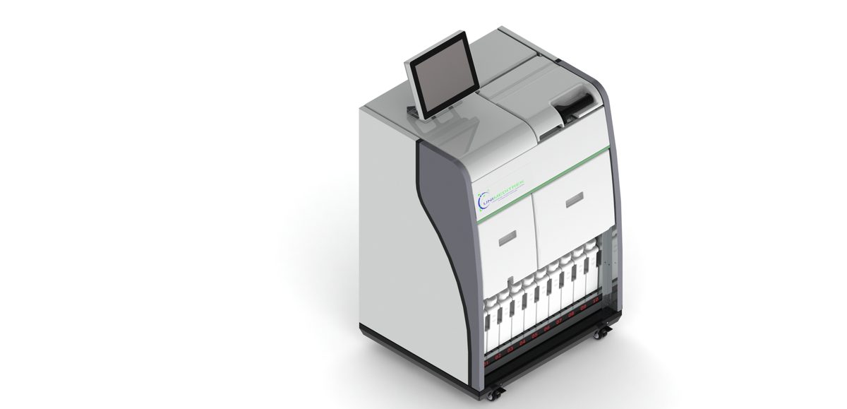
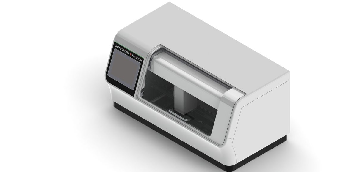
H&E is easy to do and hard to do identically every time — reagents age, dip times drift between operators, and the result wanders. That batch-to-batch consistency is exactly what an automatic slide staining machine is for: it holds the same sequence and timing on every slide. For the common failures and fixes, see troubleshooting weak or uneven H&E, and the wider picture in the histopathology workflow.
FAQs
What is hematoxylin and eosin (H&E) staining used for?
It is the routine stain in histopathology. Haematoxylin stains nuclei blue-purple and eosin stains cytoplasm and connective tissue pink, giving the contrast a pathologist needs to read tissue architecture and make most diagnoses.
What does hematoxylin stain and what does eosin stain?
Haematoxylin (basic) stains acidic structures such as the nucleus blue-purple; eosin (acidic) stains basic structures such as cytoplasm and collagen pink.
Why is H&E staining consistency difficult?
The method is simple but sensitive to reagent age and dip-time differences between operators, so results drift. Automated staining holds the same protocol on every slide for reproducible results.
Prepared and reviewed by the Unimeditrek technical team based on laboratory practice, installation and service experience. This document explains histopathology workflow, laboratory practice and equipment based on the questions we hear from hospitals, medical colleges and diagnostic laboratories — vendor-neutral where it explains the science and practical where it explains equipment.
Prepared by Unimeditrek Pvt. Ltd.. For product specifications and quotations, contact our team.