Home › Knowledge › Troubleshooting Weak or Uneven H&E Staining
Knowledge articleTroubleshooting Weak or Uneven H&E Staining
A guide for effective staining in histopathology labs
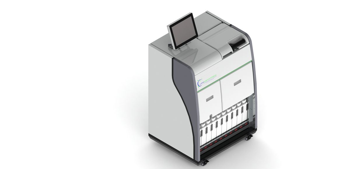
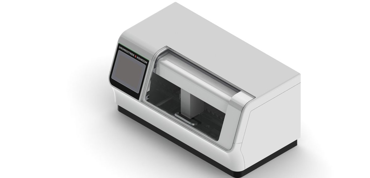
 Automatic Slide Staining Machine — Unimeditrek" style="max-width:100%;border-radius:10px"/>
Understanding H&E Staining
Hematoxylin and eosin (H&E) staining is a cornerstone technique in histopathology, enabling pathologists to visualize cellular structures and tissue architecture. Hematoxylin stains cell nuclei blue, while eosin provides a pink hue to the cytoplasm and extracellular components. The process, although seemingly straightforward, can sometimes yield weak or uneven staining results. Understanding the underlying principles of the staining process is crucial for troubleshooting these issues effectively.
When using an automatic slide staining machine, several factors can influence the staining quality, including reagent quality, staining times, and the condition of the tissues being processed. A consistent staining protocol is essential, as even minor variations can lead to significant differences in staining outcomes. It is important to familiarize yourself with the machine's settings and the specific requirements of your histological samples to achieve optimal results.
Common Causes of Weak Staining
Weak H&E staining can be attributed to several factors, the most common being inadequate hematoxylin or eosin application. If the hematoxylin solution is old or improperly prepared, it may not adequately stain the nuclei, leading to pale or weak staining. Similarly, if the eosin solution is too diluted or has been contaminated, the pink staining of the cytoplasm may be insufficient. Regularly checking the age and preparation of your staining reagents is essential for maintaining staining quality.
Another common cause of weak staining is insufficient fixation or processing of the tissue samples. If the tissue is under-fixed, the cellular components may not be adequately preserved, resulting in poor staining. Conversely, over-fixing can mask antigenic sites, also leading to weak staining. Ensuring that your automatic slide staining machine is calibrated correctly and that the fixation protocols are followed meticulously will help mitigate these issues.
Addressing Uneven Staining
Uneven H&E staining can be particularly problematic, as it may lead to misinterpretation of tissue morphology. One of the primary reasons for uneven staining is the inconsistent application of reagents, which can occur if the automatic slide staining machine is not functioning properly. Regular maintenance and calibration of the machine are crucial to ensure that it dispenses reagents evenly across all slides.
Another factor that can contribute to uneven staining is the quality of the tissue sections. If the sections are cut unevenly or if there are variations in thickness, it can lead to inconsistent staining. It is essential to ensure that the microtome is well-maintained and that the sections are cut uniformly. Additionally, the mounting medium used can affect staining quality; using a consistent medium that is compatible with the staining process will help achieve uniform results.
Optimizing Staining Protocols
To enhance the quality of H&E staining, it is beneficial to optimize the staining protocols used in your laboratory. This includes adjusting staining times, reagent concentrations, and the order of application. For instance, extending the staining time in hematoxylin can improve nuclear staining intensity, while adjusting eosin concentrations can enhance cytoplasmic staining. Conducting regular quality control checks and comparing results can help identify the most effective staining parameters for your specific samples.
It is also advisable to maintain a detailed log of staining results and any adjustments made to the protocol. This documentation can serve as a valuable reference for troubleshooting future staining issues and can help standardize processes within the lab. Training all personnel involved in the staining process to follow these optimized protocols will ensure consistency and reliability in staining outcomes.
Practical Takeaway
In summary, troubleshooting weak or uneven H&E staining requires a systematic approach. Regularly check the quality of your reagents, ensure proper fixation and processing of tissues, and maintain your automatic slide staining machine to achieve optimal results. By optimizing your staining protocols and documenting your processes, you can enhance the quality of your histopathological evaluations and minimize staining-related discrepancies.
FAQs
What should I do if the hematoxylin stain appears weak?
If hematoxylin staining is weak, check the age and preparation of the hematoxylin solution. Ensure it is fresh and properly mixed, and consider extending the staining time for better results.
How can I prevent uneven staining in my samples?
To prevent uneven staining, ensure that your automatic slide staining machine is calibrated correctly and regularly maintained. Additionally, ensure that tissue sections are cut uniformly and that the reagents are applied evenly.
What are the best practices for maintaining staining quality?
Best practices for maintaining staining quality include regularly checking reagent quality, optimizing staining protocols, documenting results, and ensuring consistent training for all personnel involved in the staining process.
Prepared and reviewed by the Unimeditrek technical team based on laboratory practice, installation and service experience. This document explains histopathology workflow, laboratory practice and equipment based on the questions we hear from hospitals, medical colleges and diagnostic laboratories — vendor-neutral where it explains the science and practical where it explains equipment.
Prepared by Unimeditrek Pvt. Ltd.. For product specifications and quotations, contact our team.