Home › Knowledge › What Is Histopathology? The Complete Workflow Explained
Knowledge articleWhat Is Histopathology? The Complete Workflow Explained
From specimen to signed-out report — every step of the histopathology pipeline.
When a surgeon removes a lump or a radiologist takes a biopsy, the tissue doesn't diagnose itself. It travels to the histopathology lab, where — over a day or so and half a dozen careful steps — it becomes a stained glass slide a pathologist can read. This is a plain walk through that journey, step by step, for anyone new to the field.
So what is histopathology?
Break the word in half: "histo" is tissue, "pathology" is the study of disease. Put together, histopathology is the study of how disease changes the microscopic structure of tissue — and it's the basis for confirming most cancers and a great many other conditions.
The workflow, start to finish
1. Receipt and grossing
The specimen is logged, described and cut down (grossed) into cassettes small enough to process. Careful labelling here is what protects a patient's identity through every step that follows.
2. Fixation
The tissue is preserved — usually in 10% neutral buffered formalin — to stop it decaying and to hold its structure. Everything downstream leans on this being done properly.
3. Processing
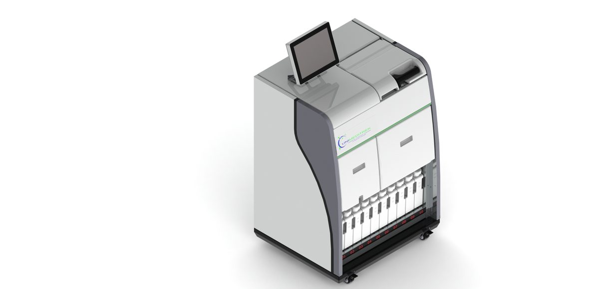
An automatic tissue processor swaps the water in the tissue for paraffin wax, so it firms up enough to section. The full story is in the complete tissue processing guide.
4. Embedding
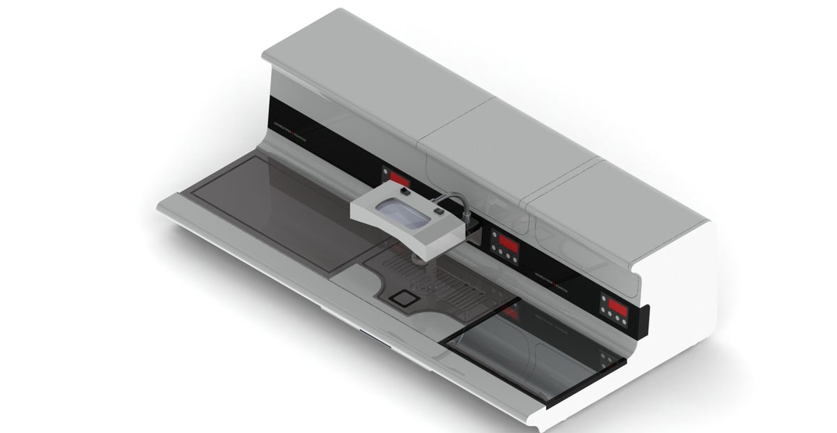
The processed tissue is set into a paraffin block at the embedding station, oriented so it'll cut the right way.
5. Microtomy
A microtome shaves the block into ribbons just microns thick. They're floated flat on a floatation bath, picked up on slides and dried on a warming table. If you're curious which instrument does what, see types of microtome.
6. Staining
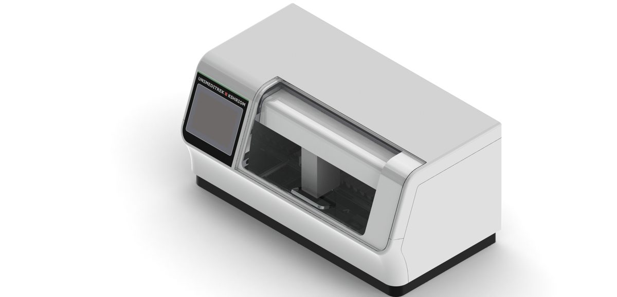
Those sections are colourless, so they're stained — routinely with H&E — on an automatic slide staining machine, which brings the structures into view.
7. Microscopy and reporting
Finally the pathologist reads the slide and writes the report, calling on special stains, immunohistochemistry or a frozen section where the case needs them.
Why the whole chain matters
Histopathology is a relay, not a sprint — a weak handoff anywhere (poor fixation, the wrong schedule, uneven staining) turns up on the final slide. That's exactly why standard equipment and protocols at every stage are worth the fuss.
FAQs
What is histopathology in simple terms?
Histopathology is the study of disease in tissue under a microscope. The lab turns a biopsy or surgical specimen into a stained slide that a pathologist reads to make a diagnosis.
What are the steps of the histopathology workflow?
Grossing, fixation, tissue processing, embedding, microtomy (sectioning), staining, and microscopy/reporting — sometimes with special stains, IHC or frozen sections added.
Prepared and reviewed by the Unimeditrek technical team based on laboratory practice, installation and service experience. This document explains histopathology workflow, laboratory practice and equipment based on the questions we hear from hospitals, medical colleges and diagnostic laboratories — vendor-neutral where it explains the science and practical where it explains equipment.
Prepared by Unimeditrek Pvt. Ltd.. For product specifications and quotations, contact our team.