Home › Knowledge › Histopathology Notes for MLT & DMLT Students
Knowledge articleHistopathology Notes for MLT & DMLT Students
The techniques, reagents and steps you need to know — in one place.
If you're studying MLT or DMLT, histopathology can feel like a pile of reagents and steps to memorise. It's easier than that once you see it as one journey — tissue in, stained slide out. These notes lay out that journey, the reagents and kit at each stage, and the points examiners love to ask. Follow the links when you want the deeper version.
Learn this order first
Grossing → Fixation → Processing (dehydration → clearing → infiltration) → Embedding → Microtomy → Staining → Microscopy. Get that sequence solid and everything else hangs off it — the full explanation is in what is histopathology.
Reagents, and what each one is for
It's much easier to remember reagents by their job than as a list. Formalin (10% neutral buffered) fixes — it preserves. Graded alcohols dehydrate — they pull water out. Xylene (or a substitute) clears — it's the go-between that mixes with wax. Paraffin wax does the infiltration and embedding. And haematoxylin & eosin is the routine stain — nuclei blue, cytoplasm pink.
Equipment to picture
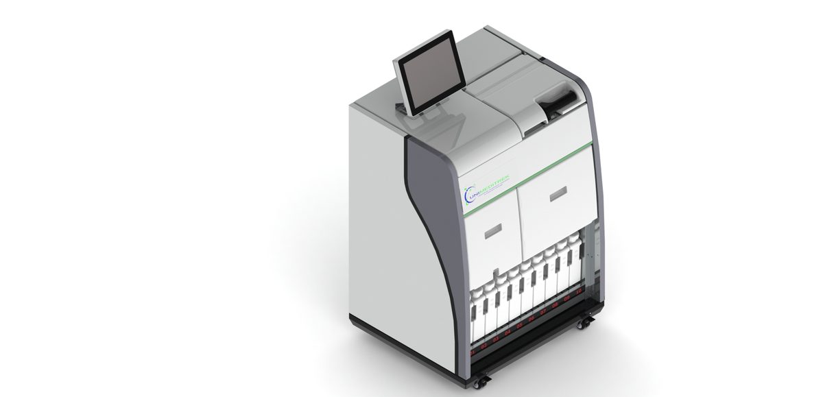
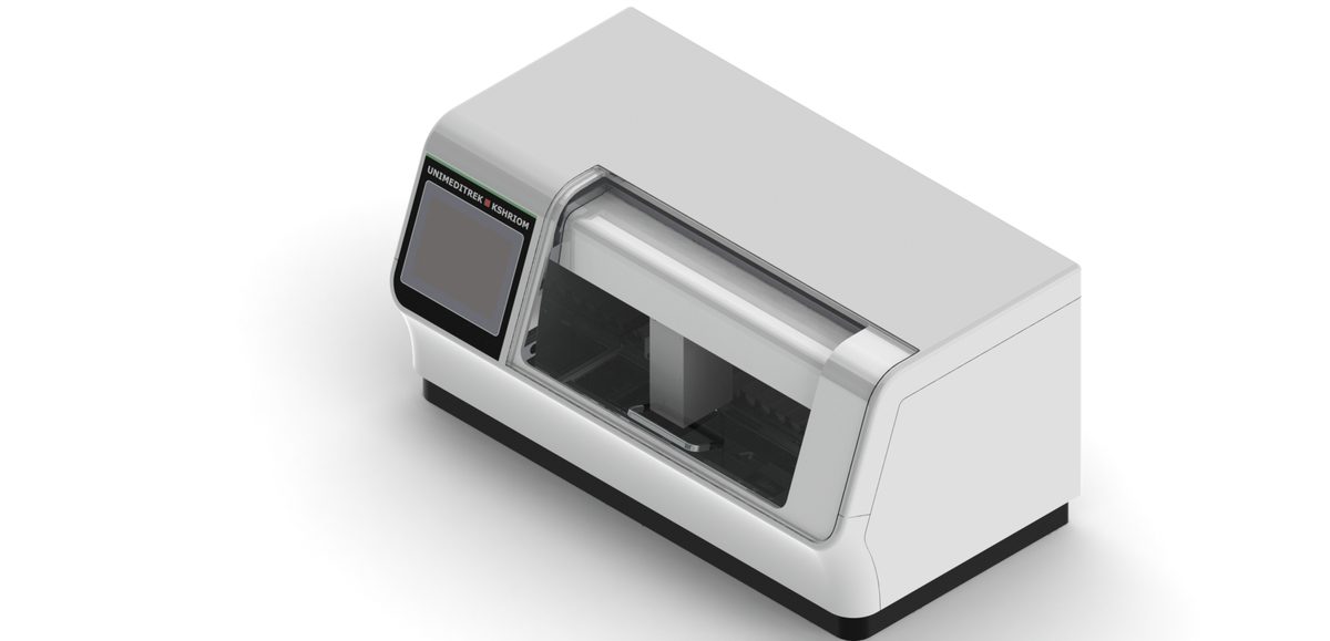
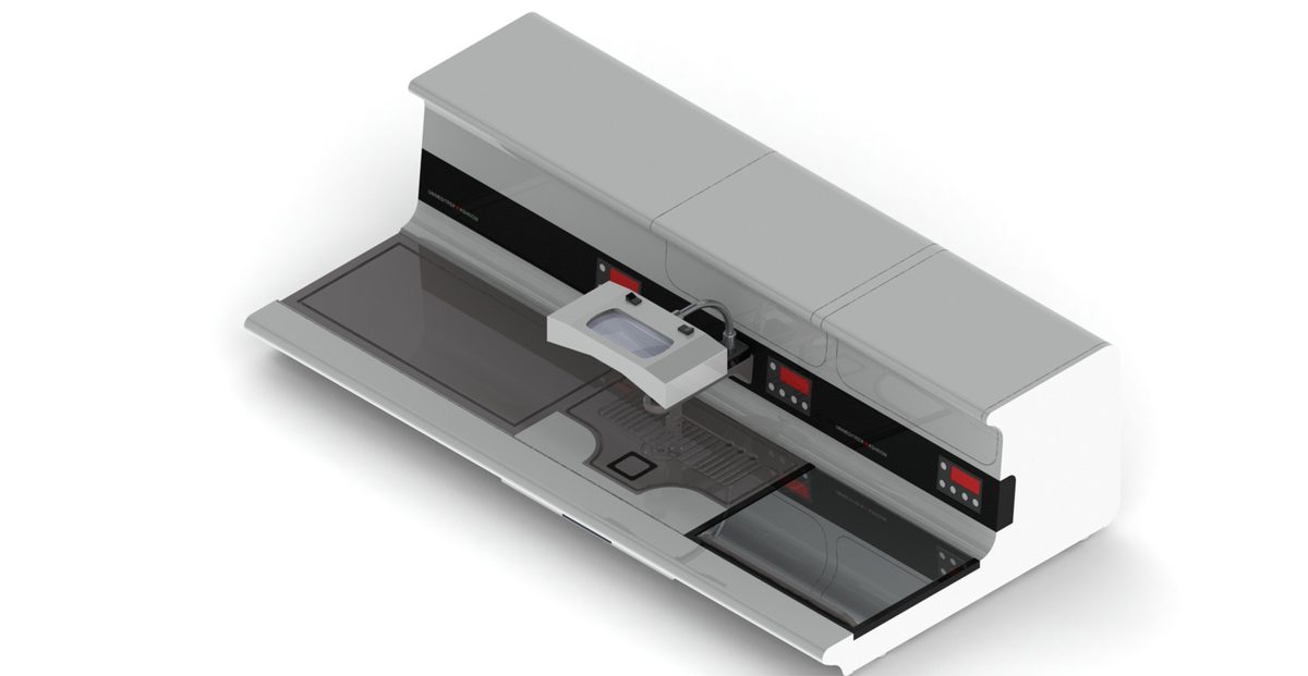
A tissue processor automates dehydration, clearing and infiltration; an embedding station sets the blocks; a microtome cuts them (know your types of microtome); a floatation bath and warming table flatten and dry the sections; and a slide staining machine automates the staining.
The questions that come up
Expect to be asked the order of the processing steps and why each is done; why fixation has to be adequate before processing; the usual thickness of routine paraffin sections (around 3–5 microns); what H&E stains and the colours you'd expect; and the difference between a frozen section (cryostat, rapid) and a routine paraffin one. Learn each stage from the linked guides and you'll have both the practical technique and the theory to back it up.
FAQs
What are the main histopathology techniques for MLT students?
Fixation, tissue processing (dehydration, clearing, infiltration), embedding, microtomy, and staining (routinely H&E), followed by microscopy. Each has specific reagents and equipment.
What thickness are routine paraffin sections cut at?
Routine paraffin sections are typically cut at around 3–5 microns on a rotary microtome for H&E staining.
Prepared and reviewed by the Unimeditrek technical team based on laboratory practice, installation and service experience. This document explains histopathology workflow, laboratory practice and equipment based on the questions we hear from hospitals, medical colleges and diagnostic laboratories — vendor-neutral where it explains the science and practical where it explains equipment.
Prepared by Unimeditrek Pvt. Ltd.. For product specifications and quotations, contact our team.