Home › Knowledge › Cell Block Preparation in Cytology: Methods for Histology-Grade Material
Knowledge articleCell Block Preparation in Cytology: Methods for Histology-Grade Material
Turning aspirates and effusions into paraffin blocks you can stain and do IHC on.
There's a quiet revival happening in cytology, and it's the humble cell block. A cell block takes material that would otherwise only give you smears — an aspirate, an effusion, a washing — and turns it into a paraffin block you can section and stain like any biopsy. That one move unlocks a full IHC panel, special stains and molecular work on samples that used to be a diagnostic dead end. Here's how it's done, and where it tends to go wrong.
Why bother making one
Three reasons keep coming up: you can run a proper IHC panel to pin down a diagnosis on limited material; you get reproducible sections for special stains and molecular tests; and you end up with a permanent FFPE block to archive. In an age of ever-smaller samples, that's not a luxury.
How it's done
Every method is really the same idea — get the cells into a compact, handleable pellet before processing. The plasma-thrombin approach clots them into a firm pellet; cell/agar or HistoGel-type methods set the pellet in a gel disc; and at its simplest, a spun-down sediment is fixed and wrapped for the cassette. Use whichever your lab is set up for and can do consistently.
Processing and embedding
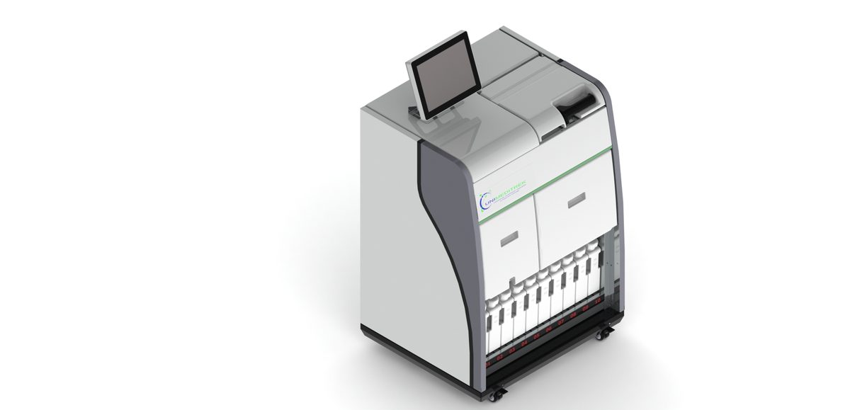
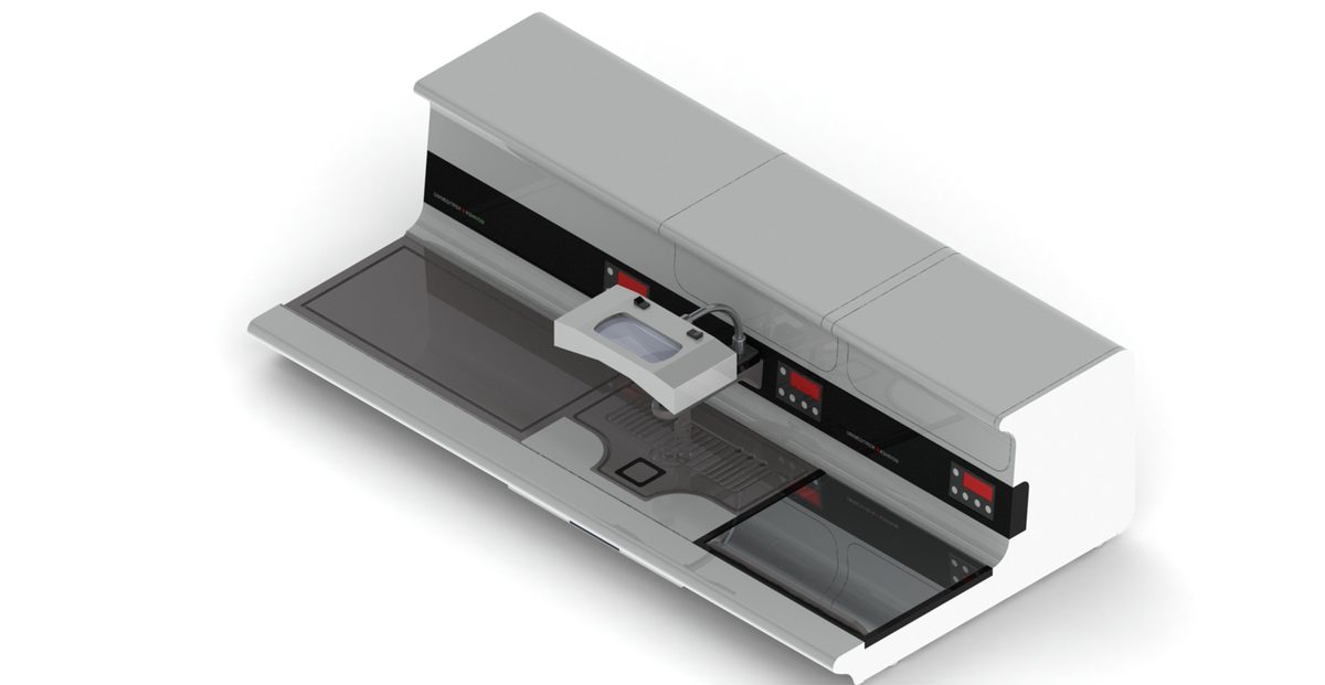
The pellet is small, so treat it like a biopsy — brief fixation, a short schedule (see protocols), then embed it flat and central at the embedding station so you don't face straight past the cells.
If the block comes out sparse
"There aren't enough cells" is the classic complaint, and it's nearly always a concentration problem, not a sectioning one. Maximise the pellet, colour it so you can see it, embed it in one plane — and the H&E and IHC read as cleanly as they do on tissue.
FAQs
What is a cell block in cytology?
A cell block is a paraffin block made from concentrated cytology material (aspirates, effusions, washings) so it can be sectioned and stained like a tissue biopsy, enabling IHC, special stains and molecular tests.
Why are cell blocks useful for IHC?
They provide reproducible FFPE sections from limited cytology samples, so a full immunohistochemistry panel can be run to refine a diagnosis when smears alone are not enough.
Prepared and reviewed by the Unimeditrek technical team based on laboratory practice, installation and service experience. This document explains histopathology workflow, laboratory practice and equipment based on the questions we hear from hospitals, medical colleges and diagnostic laboratories — vendor-neutral where it explains the science and practical where it explains equipment.
Prepared by Unimeditrek Pvt. Ltd.. For product specifications and quotations, contact our team.