Home › Knowledge › Why Stable Temperature Prevents Folds and Tears
Knowledge articleWhy Stable Temperature Prevents Folds and Tears
Understanding the Importance in Histopathology
The Role of Temperature in Histopathology
In histopathology, the preparation of tissue samples is a delicate process that requires precision and care. One of the critical factors influencing the quality of tissue sectioning is temperature stability. When tissues are prepared for microscopic examination, they undergo various processes, including fixation, embedding, and sectioning. Each of these steps demands a controlled environment to preserve the integrity of the samples. Fluctuations in temperature can lead to significant issues, including folds and tears in the tissue sections, which can compromise the diagnostic quality of the specimens.
A stable temperature is essential for maintaining the physical properties of the tissue. For example, paraffin-embedded tissues must be kept at a consistent temperature to ensure that they remain pliable and easy to cut. If the temperature fluctuates, the paraffin can become too hard or too soft, leading to uneven sectioning and potential damage to the tissue. This can result in folds, tears, or even complete loss of diagnostic material, ultimately affecting patient care.
The Mechanism Behind Temperature-Induced Damage
Temperature changes can alter the viscosity of embedding media such as paraffin wax. When the temperature is too high, the wax can become overly soft, making it difficult to achieve clean cuts. Conversely, if the temperature is too low, the wax can harden excessively, leading to brittle sections that are prone to tearing. This mechanical stress on the tissue can create artifacts that obscure important histological features, which are crucial for accurate diagnosis.
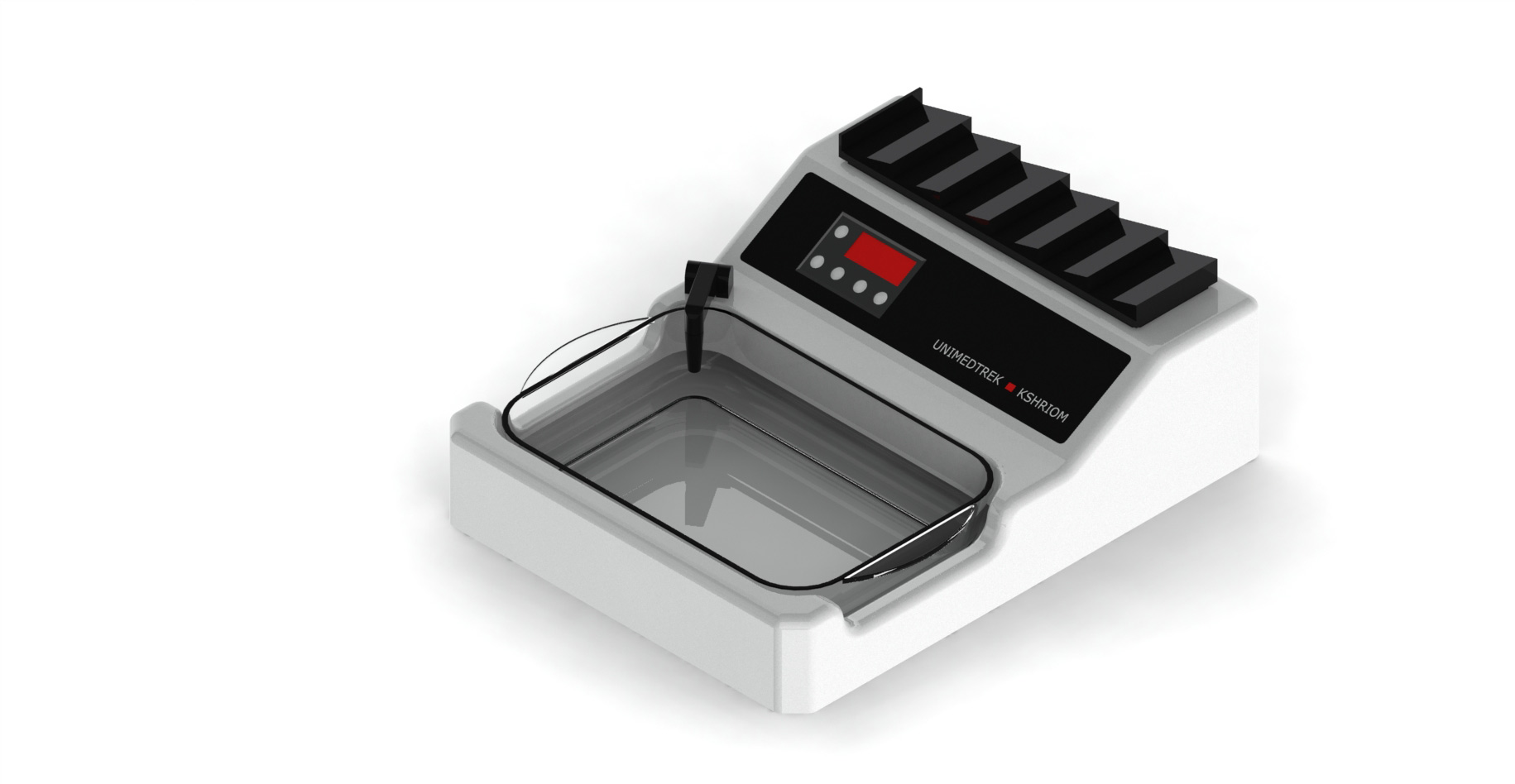
Furthermore, temperature fluctuations can also impact the hydration state of the tissue. Tissues that are not adequately hydrated may shrink or expand unevenly when subjected to varying temperatures. This can lead to distortion in the morphology of the cells and tissues, making it challenging for pathologists to interpret the samples accurately. The use of a digital tissue floatation bath, which maintains a stable temperature, can mitigate these risks by providing a controlled environment for tissue processing.
Benefits of Using a Digital Tissue Floatation Bath
A digital tissue floatation bath offers several advantages over traditional methods of temperature control in histopathology. One of the primary benefits is the precision that digital technology provides. Pathologists and lab technicians can set and monitor the temperature with high accuracy, ensuring that the tissue remains at the optimal temperature throughout the sectioning process. This precision reduces the likelihood of temperature-induced artifacts and enhances the overall quality of the tissue sections.
Additionally, digital floatation baths often come equipped with features such as programmable settings and alarms, which can further enhance workflow efficiency. For instance, technicians can pre-set the bath to the desired temperature before starting their work, allowing for a seamless transition into the sectioning phase. This not only saves time but also ensures that the samples are treated consistently, contributing to reproducible results in histopathological assessments.
Best Practices for Using a Digital Tissue Floatation Bath
To maximize the benefits of a digital tissue floatation bath, it is essential to follow best practices during its use. First, always calibrate the device according to the manufacturer's instructions to ensure accurate temperature readings. Regular maintenance and cleaning of the bath will also help prevent contamination and ensure optimal performance.
Moreover, it is advisable to monitor the temperature regularly, especially during busy periods when multiple samples are being processed. Keeping a log of temperature readings can help identify any anomalies that may arise, allowing for timely adjustments. Additionally, using high-quality embedding media and ensuring proper hydration of tissues prior to sectioning can further enhance the quality of the final specimens.
Conclusion and Practical Takeaway
In summary, maintaining a stable temperature is crucial for preventing folds and tears in tissue sections during histopathological preparation. The use of a digital tissue floatation bath can significantly improve the quality of tissue processing by providing a controlled environment that minimizes temperature fluctuations. By adhering to best practices, histopathology labs can ensure that their samples are preserved with integrity, ultimately leading to more accurate diagnoses and better patient outcomes.
As a practical takeaway, ensure that your digital tissue floatation bath is calibrated correctly and regularly maintained. Implementing a systematic approach to temperature monitoring and sample handling can greatly enhance the quality of your histopathological work.
FAQs
What is the ideal temperature for a tissue floatation bath?
The ideal temperature for a tissue floatation bath typically ranges between 40°C to 60°C, depending on the type of embedding medium used.
How often should I calibrate my digital tissue floatation bath?
It is advisable to calibrate your digital tissue floatation bath at regular intervals, such as monthly or quarterly, to ensure accurate temperature readings.
Can temperature fluctuations affect tissue staining results?
Yes, temperature fluctuations can lead to artifacts in tissue sections, which may obscure histological details and affect staining results.
Prepared and reviewed by the Unimeditrek technical team based on laboratory practice, installation and service experience. This document explains histopathology workflow, laboratory practice and equipment based on the questions we hear from hospitals, medical colleges and diagnostic laboratories — vendor-neutral where it explains the science and practical where it explains equipment.
Prepared by Unimeditrek Pvt. Ltd.. For product specifications and quotations, contact our team.