Home › Knowledge › Why Fixation Quality Decides Everything Downstream
Knowledge articleWhy Fixation Quality Decides Everything Downstream
Understanding the Importance of Fixation in Histopathology
The Role of Fixation in Histopathology
Fixation is a critical step in the histopathological workflow, serving as the foundation for all subsequent processes. The primary purpose of fixation is to preserve tissue morphology and prevent autolysis and putrefaction. By stabilizing the cellular structure, fixation allows for accurate examination and diagnosis. Without proper fixation, the integrity of the tissue can be compromised, leading to misleading results during microscopic evaluation.
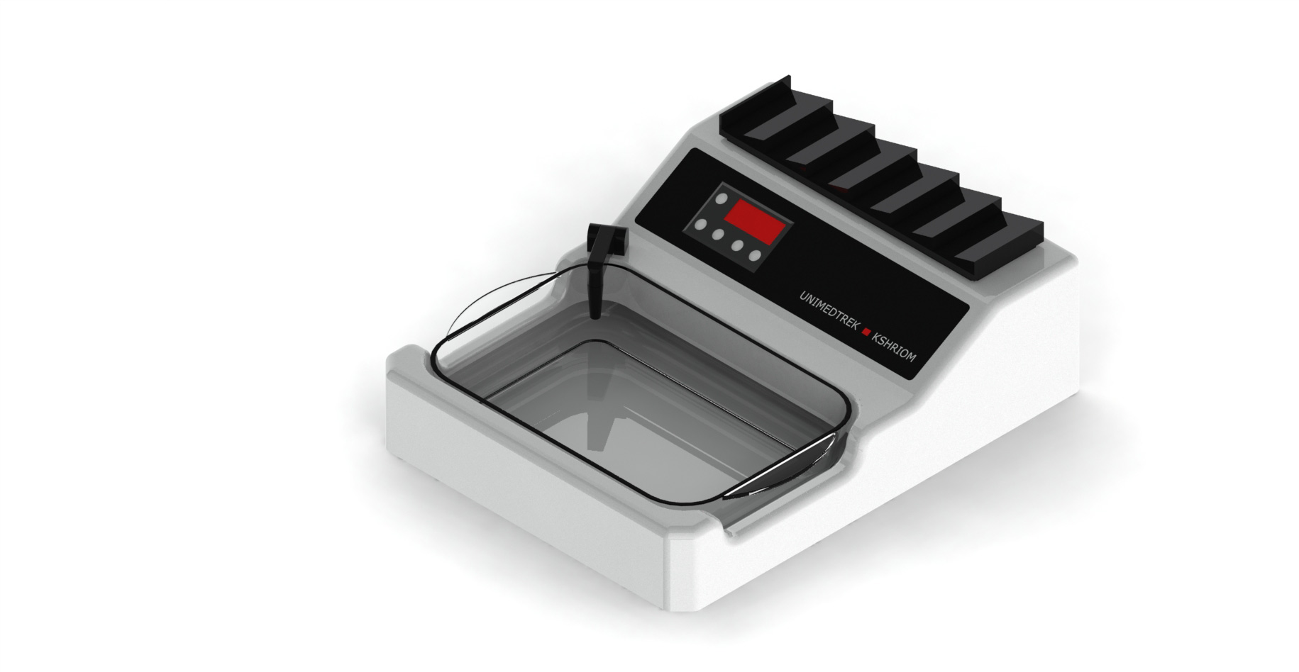
Different fixation techniques can yield varying results, and the choice of fixative can greatly influence the quality of the tissue sample. For instance, formalin is a commonly used fixative that penetrates tissues effectively, yet its fixation time and concentration can affect the ultimate outcome. In this context, employing a digital tissue floatation bath can enhance the quality of tissue processing by ensuring that samples are adequately floated and oriented for optimal embedding.
Impact of Fixation on Staining Techniques
The fixation quality directly impacts the staining techniques employed in histopathology. Properly fixed tissues are essential for achieving optimal staining results, as fixation preserves cellular antigens and structures that stains target. If fixation is inadequate, it may lead to poor staining quality, which can obscure diagnostic features and complicate interpretation.
For example, immunohistochemical staining relies heavily on the preservation of antigenicity, which can be compromised by suboptimal fixation. As a result, pathologists may miss critical diagnostic markers, leading to misdiagnosis or delayed treatment. The digital tissue floatation bath plays a pivotal role in this process by providing an environment that ensures even distribution of tissue sections, promoting better interaction with staining reagents.
Consequences of Poor Fixation
When fixation is not performed correctly, the consequences can ripple throughout the histopathological workflow. Poor fixation can lead to artifacts, such as shrinkage or distortion of tissue samples, which can misrepresent the underlying pathology. These artifacts can confuse pathologists and lead to erroneous conclusions about the disease state.
Moreover, inadequate fixation can result in the loss of important cellular details that are critical for accurate diagnosis. This can necessitate repeat biopsies or additional testing, which not only increases the burden on patients but also strains laboratory resources. The importance of a reliable digital tissue floatation bath cannot be overstated, as it helps ensure that samples are handled gently and accurately, reducing the risk of artifacts during the embedding process.
Optimizing Fixation Protocols
To maximize the quality of fixation, laboratories should establish and follow standardized fixation protocols. This includes determining the appropriate fixation time and fixative concentration based on the type of tissue being processed. Additionally, it is essential to ensure that tissues are fixed promptly after excision to prevent degradation.
Incorporating a digital tissue floatation bath into the workflow can further enhance fixation quality. This equipment provides a controlled environment for tissue sections, allowing for optimal orientation and reducing the likelihood of damage during the transfer to embedding molds. By prioritizing fixation quality, labs can improve the overall accuracy of their histopathological assessments.
Practical Takeaway: Ensuring Quality Control
Quality control is paramount in histopathology, particularly concerning fixation. Laboratories should implement regular training sessions for technicians to reinforce the importance of proper fixation techniques. Additionally, routine audits of fixation processes can help identify areas for improvement.
Utilizing advanced equipment, such as a digital tissue floatation bath, can contribute to maintaining high standards of fixation quality. By focusing on these key areas, histopathology labs can enhance their diagnostic capabilities and ultimately improve patient outcomes.
Further reading
For peer-reviewed depth on histopathology practice and quality, these leading journals are excellent references:
- The Journal of Pathology
- Histopathology (Wiley)
- Modern Pathology (USCAP)
- Archives of Pathology & Laboratory Medicine (CAP)
- American Journal of Clinical Pathology
- Journal of Clinical Pathology (BMJ)
Tissue Floatation Bath — engineered and serviced in India
Unimeditrek manufactures the Tissue Floatation Bath with pan-India installation and service. Explore the full specifications, request a demo, or register to download the brochure — direct from the manufacturer.
View Tissue Floatation Bath & request a quote →FAQs
What is the best fixative for tissue samples?
The choice of fixative depends on the type of tissue and the downstream applications. Formalin is commonly used, but alternatives like glutaraldehyde may be suitable for specific needs.
How does a digital tissue floatation bath improve fixation?
A digital tissue floatation bath provides a controlled environment for tissue sections, ensuring proper orientation and reducing the risk of artifacts during embedding.
What are the signs of poor fixation in histopathology?
Signs of poor fixation include tissue shrinkage, distortion, and artifacts that can obscure cellular details, leading to misdiagnosis.
Prepared and reviewed by the Unimeditrek technical team based on laboratory practice, installation and service experience. This document explains histopathology workflow, laboratory practice and equipment based on the questions we hear from hospitals, medical colleges and diagnostic laboratories — vendor-neutral where it explains the science and practical where it explains equipment.
Prepared by Unimeditrek Pvt. Ltd.. For product specifications and quotations, contact our team.