Home › Knowledge › Why Fixation Quality Decides Everything Downstream
Knowledge articleWhy Fixation Quality Decides Everything Downstream
Understanding the critical role of fixation in histopathology
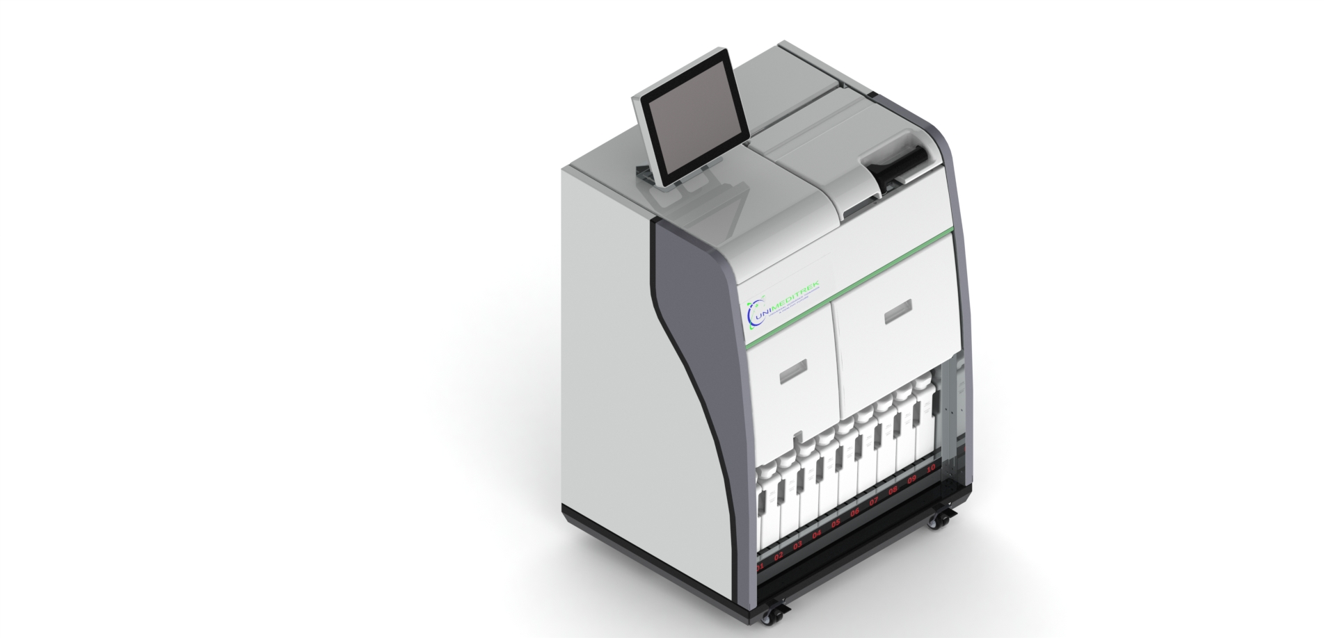
 Tissue Embedding Station — Unimeditrek" style="max-width:100%;border-radius:10px"/>
The Importance of Fixation in Histopathology
In histopathology, the fixation process is the cornerstone of tissue preservation. It involves the treatment of biological tissues with fixatives to prevent autolysis and decay, allowing for accurate examination under a microscope. The quality of fixation directly impacts the morphology and cellular detail of the tissue, which are essential for pathologists to make informed diagnoses. Poor fixation can lead to artifacts, distortion, or even loss of cellular structures, ultimately compromising the integrity of the histological analysis.
When tissues are not properly fixed, the consequences can be severe. For instance, if the fixation is inadequate, it can result in the loss of antigenicity, making immunohistochemical staining ineffective. This means that important markers may not be visualized, leading to misinterpretation of the disease state. Thus, the fixation process is not merely a preparatory step; it is a critical determinant of the quality of downstream procedures, including embedding, sectioning, and staining.
Types of Fixatives and Their Impact
Different types of fixatives serve distinct purposes and can significantly influence the outcome of histological examinations. Formaldehyde, for example, is the most commonly used fixative in histopathology due to its ability to preserve cellular morphology while cross-linking proteins. However, its effectiveness can be compromised if tissues are not immersed in the fixative for an adequate duration or if the fixative is not fresh.
Other fixatives, such as glutaraldehyde, are often used for electron microscopy due to their superior preservation of ultrastructural details. Understanding the properties of each fixative and the specific requirements of the tissue being processed is essential for histotechnologists. The choice of fixative can affect the compatibility with subsequent procedures, including those performed in a paraffin embedding station, thereby influencing the overall quality of the diagnostic output.
Fixation Time and Temperature Considerations
The time and temperature at which fixation occurs are critical parameters that can greatly affect tissue quality. Insufficient fixation time can result in incomplete penetration of the fixative, leading to areas of necrosis or inadequate preservation of cellular components. Conversely, over-fixation can cause excessive cross-linking and hardening of the tissue, making it difficult to cut thin sections during the embedding process.
Temperature also plays a vital role in fixation. Most fixatives are more effective at room temperature, while cold temperatures can slow down the fixation process. It is crucial for laboratory personnel to adhere to recommended fixation protocols, as deviations can lead to significant variations in tissue quality. Proper training and adherence to fixation guidelines are essential to ensure that the tissue is optimally preserved for downstream applications.
The Role of the Paraffin Embedding Station
A paraffin embedding station is an essential piece of equipment in histopathology labs, particularly in the context of tissue processing. After fixation, tissues are dehydrated and infiltrated with paraffin wax, which provides structural support for thin sectioning. The quality of the embedding process is highly dependent on the quality of fixation. If the tissue is not well-fixed, it may not adequately infiltrate with paraffin, leading to poor quality sections that are difficult to cut and stain.
Moreover, a well-designed paraffin embedding station can enhance workflow efficiency, allowing for better handling of multiple samples simultaneously. This is particularly important in busy laboratories where time is of the essence. By ensuring that the embedding process is carried out smoothly and efficiently, labs can minimize the risk of errors and improve the overall quality of the histological specimens produced.
Practical Takeaways for Histopathology Labs
For histopathology labs, understanding the importance of fixation cannot be overstated. Proper training for laboratory personnel on fixation protocols, including the choice of fixatives, fixation times, and temperature, is crucial. Regular calibration and maintenance of the paraffin embedding station are also important to ensure optimal performance.
By prioritizing fixation quality, laboratories can enhance the reliability of their histopathological results. This not only aids pathologists in making accurate diagnoses but also ultimately benefits patient care. A commitment to quality in every step of the histopathological process, from fixation to embedding, is essential for achieving the best outcomes.
Further reading
For peer-reviewed depth on histopathology practice and quality, these leading journals are excellent references:
- The Journal of Pathology
- Histopathology (Wiley)
- Modern Pathology (USCAP)
- Archives of Pathology & Laboratory Medicine (CAP)
- American Journal of Clinical Pathology
- Journal of Clinical Pathology (BMJ)
Tissue Embedding Station — engineered and serviced in India
Unimeditrek manufactures the Tissue Embedding Station with pan-India installation and service. Explore the full specifications, request a demo, or register to download the brochure — direct from the manufacturer.
View Tissue Embedding Station & request a quote →FAQs
What are the common fixatives used in histopathology?
Common fixatives include formaldehyde, glutaraldehyde, and ethanol. Each has specific properties that affect tissue preservation.
How long should tissues be fixed for optimal results?
The fixation time varies depending on the type of tissue and fixative used, but generally, tissues should be fixed for several hours to overnight for optimal preservation.
What issues can arise from poor fixation?
Poor fixation can lead to artifacts, loss of cellular detail, and ineffective staining, ultimately compromising diagnostic accuracy.
Prepared and reviewed by the Unimeditrek technical team based on laboratory practice, installation and service experience. This document explains histopathology workflow, laboratory practice and equipment based on the questions we hear from hospitals, medical colleges and diagnostic laboratories — vendor-neutral where it explains the science and practical where it explains equipment.
Prepared by Unimeditrek Pvt. Ltd.. For product specifications and quotations, contact our team.