Home › Knowledge › Tissue Fixation in Histopathology: Formalin, Timing, Pitfalls
Knowledge articleTissue Fixation in Histopathology: Formalin, Timing, Pitfalls
Understanding the essentials of tissue fixation
Introduction to Tissue Fixation
Tissue fixation is a critical step in histopathology that preserves cellular structure and integrity, allowing for accurate diagnosis. The most commonly used fixative is formalin, which penetrates tissues and cross-links proteins, ensuring that cellular morphology is maintained during subsequent processing.
The Role of Formalin in Tissue Fixation
Formalin, typically a 10% solution of formaldehyde, is favored due to its effectiveness in preserving tissue architecture. It is essential to use fresh formalin and ensure that the fixation process begins promptly after tissue collection to prevent autolysis and decay. The ideal fixation time can vary, but generally, a minimum of 6-8 hours is recommended for optimal results.
Timing: The Key to Effective Fixation
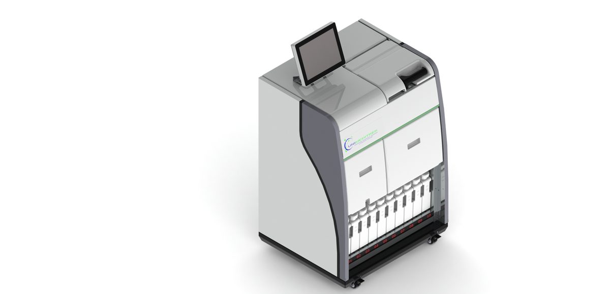
Timing in tissue fixation cannot be overstated. Under-fixation may lead to poor morphological preservation, while over-fixation can obscure cellular details. For most tissues, a fixation time of 24 hours is optimal, but this can vary based on tissue type and size. For larger specimens, it is advisable to use an Automatic Vacuum Tissue Processor to ensure uniform fixation throughout the tissue.
Common Pitfalls in Tissue Fixation
Several common pitfalls can compromise tissue fixation:
- Inadequate Fixation Time: Not allowing sufficient time for formalin to penetrate the tissue can lead to incomplete fixation.
- Improper Tissue Size: Large tissue samples can result in uneven fixation; cutting them into smaller pieces can improve penetration.
- Temperature Effects: Fixation should occur at room temperature; extreme temperatures can alter the fixation process.
- Storage Conditions: Storing fixed tissues in formalin for extended periods can lead to over-fixation.
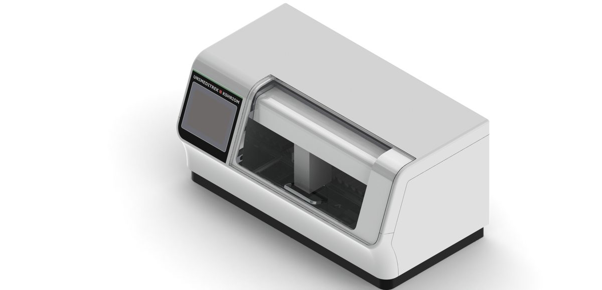
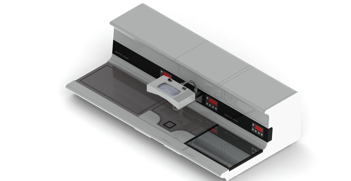
Utilizing a Tissue Embedding Station and a Digital Tissue Floatation Bath can help streamline the embedding process and ensure that tissues are adequately prepared for sectioning.
Conclusion and Practical Takeaway
In summary, proper tissue fixation is essential for accurate histopathological analysis. By understanding the role of formalin, adhering to optimal timing, and avoiding common pitfalls, laboratories can enhance the quality of their histological preparations. To further improve workflow, consider integrating equipment such as the Automatic Slide Staining Machine and Cryostat Microtome to achieve consistent and reliable results.
FAQs
What is the purpose of tissue fixation in histopathology?
Tissue fixation preserves cellular structure and morphology, allowing for accurate diagnosis in histopathological analysis.
How long should tissues be fixed in formalin?
Generally, tissues should be fixed in formalin for a minimum of 6-8 hours, with 24 hours being optimal for most specimens.
Prepared and reviewed by the Unimeditrek technical team based on laboratory practice, installation and service experience. This document explains histopathology workflow, laboratory practice and equipment based on the questions we hear from hospitals, medical colleges and diagnostic laboratories — vendor-neutral where it explains the science and practical where it explains equipment.
Prepared by Unimeditrek Pvt. Ltd.. For product specifications and quotations, contact our team.