Home › Knowledge › Special Stains: When and Why They Earn Their Place
Knowledge articleSpecial Stains: When and Why They Earn Their Place
Understanding the Role of Special Stains in Histopathology
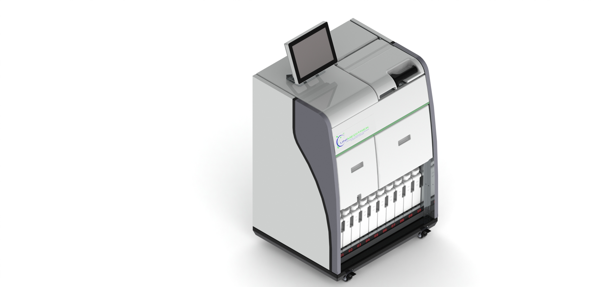
 Automatic Slide Staining Machine — Unimeditrek" style="max-width:100%;border-radius:10px"/>
Understanding Special Stains
Special stains are crucial tools in the histopathology laboratory, utilized to highlight specific cellular components or tissue structures that may not be discernible with routine stains. Unlike standard stains such as Hematoxylin and Eosin (H&E), which provide a general overview of tissue architecture, special stains are designed to target particular elements, allowing pathologists to conduct a more detailed analysis. This specificity can be vital for accurate diagnosis, especially in complex cases.
The choice of a special stain depends on the clinical question at hand. For instance, if a pathologist suspects the presence of a fungal infection, they may employ a special stain like Gomori's methenamine silver (GMS) to visualize fungal cell walls. Similarly, stains such as Masson’s trichrome can differentiate between collagen and muscle fibers, which can be pivotal in diagnosing fibrosis or muscle disorders. The ability to discern these details can significantly impact patient management and treatment decisions.
Types of Special Stains
There are numerous special stains available, each serving a unique purpose. Some of the most commonly used special stains include periodic acid-Schiff (PAS), which is effective in highlighting polysaccharides and mucosubstances; and Ziehl-Neelsen stain, which is pivotal in identifying acid-fast bacilli, such as those causing tuberculosis. Other stains, like the Oil Red O, are utilized to visualize lipids in frozen sections.
Each stain has its own methodology, requiring careful preparation and application. For example, the PAS stain involves a series of steps including oxidation, staining, and counterstaining, which must be meticulously followed to achieve optimal results. The linear slide staining machine can streamline this process, ensuring consistent results and reducing the potential for human error during staining.
The Role of Special Stains in Diagnosis
Special stains play a significant role in the diagnostic process, particularly in challenging cases where routine staining may not provide sufficient information. For example, in the evaluation of tumors, special stains can help determine the origin of the tumor based on the specific characteristics of the cells. This is particularly important in differentiating between various types of carcinomas and sarcomas, where treatment protocols may differ significantly.
Moreover, special stains can assist in identifying infectious agents, autoimmune diseases, and other pathological conditions. For instance, the use of the Giemsa stain can reveal the presence of certain parasites in tissue samples, while immunohistochemical stains can highlight the presence of specific proteins associated with malignancies. The ability to utilize these stains effectively can enhance diagnostic accuracy and ultimately improve patient outcomes.
Implementing Special Stains in the Laboratory
To effectively implement special stains in a histopathology laboratory, it is essential to have a well-structured protocol in place. This includes proper training for laboratory personnel on the specific techniques and precautions associated with each stain. Ensuring that the staining protocols are followed meticulously can greatly enhance the reliability of the results.
Additionally, the use of a linear slide staining machine can facilitate the staining process, making it more efficient and reproducible. These machines can automate many of the time-consuming steps involved in staining, allowing for greater throughput and consistency. Regular maintenance and calibration of the staining machine are also crucial to ensure optimal performance and accuracy in results.
Practical Takeaway for Histopathologists
For histopathologists and lab technicians, understanding the significance of special stains and their applications is essential for enhancing diagnostic capabilities. By integrating special stains into routine practice, along with utilizing advanced equipment like the linear slide staining machine, laboratories can improve their diagnostic accuracy and efficiency. It is advisable to stay updated on the latest staining techniques and emerging stains that may further refine diagnostic processes.
Incorporating special stains into the diagnostic workflow not only enhances the quality of histopathological evaluation but also contributes to better patient care. Continuous education and training in the use of these stains can empower laboratory professionals to make informed decisions that can significantly impact clinical outcomes.
Further reading
For peer-reviewed depth on histopathology practice and quality, these leading journals are excellent references:
- The Journal of Pathology
- Histopathology (Wiley)
- Modern Pathology (USCAP)
- Archives of Pathology & Laboratory Medicine (CAP)
- American Journal of Clinical Pathology
- Journal of Clinical Pathology (BMJ)
Automatic Slide Staining Machine — engineered and serviced in India
Unimeditrek manufactures the Automatic Slide Staining Machine with pan-India installation and service. Explore the full specifications, request a demo, or register to download the brochure — direct from the manufacturer.
View Automatic Slide Staining Machine & request a quote →FAQs
What are special stains used for in histopathology?
Special stains are used to highlight specific cellular components or tissue structures that are not visible with routine stains, aiding in accurate diagnosis.
How do special stains differ from routine stains?
Special stains target specific elements within tissues, providing detailed information, while routine stains like H&E offer a general overview of tissue architecture.
Why is the linear slide staining machine important in histopathology?
The linear slide staining machine automates the staining process, ensuring consistency, efficiency, and reducing the likelihood of human error in special stain applications.
Prepared and reviewed by the Unimeditrek technical team based on laboratory practice, installation and service experience. This document explains histopathology workflow, laboratory practice and equipment based on the questions we hear from hospitals, medical colleges and diagnostic laboratories — vendor-neutral where it explains the science and practical where it explains equipment.
Prepared by Unimeditrek Pvt. Ltd.. For product specifications and quotations, contact our team.