Home › Knowledge › Special Stains: Their Importance in Histopathology
Knowledge articleSpecial Stains: Their Importance in Histopathology
Understanding the Role of Special Stains in Tissue Analysis
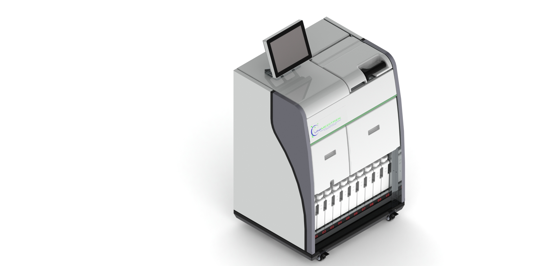
 Tissue Embedding Station — Unimeditrek" style="max-width:100%;border-radius:10px"/>
Introduction to Special Stains
In the field of histopathology, special stains have a pivotal role in enhancing the visualization of specific cellular components and structures within tissue samples. Unlike routine stains such as Hematoxylin and Eosin (H&E), which provide a general overview of tissue morphology, special stains are designed to highlight particular elements, making them indispensable in diagnosing various diseases. Their application can provide critical insights that may not be evident through standard staining techniques.
Special stains can be employed to identify a wide array of substances, including microorganisms, connective tissue components, and cellular inclusions. For instance, they can help in detecting fungal infections, identifying amyloid deposits, or differentiating between various types of connective tissue diseases. This specificity is what earns special stains their rightful place in the histopathology laboratory, allowing pathologists to make more accurate diagnoses and treatment plans.
Types of Special Stains and Their Applications
There are numerous types of special stains, each tailored to reveal distinct features of tissues. Some common special stains include the Periodic Acid-Schiff (PAS) stain, which is particularly effective in highlighting polysaccharides and mucosubstances, and the Masson's Trichrome stain, which differentiates between collagen and muscle fibers. These stains are routinely used in various clinical scenarios, such as identifying glycogen storage diseases or assessing fibrosis in liver biopsies.
Another important category of special stains includes those used for detecting microorganisms. For example, the Giemsa stain is utilized to visualize bacteria and parasites, while the Ziehl-Neelsen stain is essential for identifying acid-fast bacilli, such as Mycobacterium tuberculosis. The ability to visualize these pathogens directly within tissue sections is crucial for accurate diagnosis and subsequent management of infectious diseases.
The Process of Applying Special Stains
The application of special stains requires meticulous attention to detail and a thorough understanding of the staining protocols. Each stain has its own specific preparation and staining procedure, which may involve multiple steps, including fixation, dehydration, and rehydration of the tissue samples. The use of a tissue embedder is critical in this process, as it ensures that the tissues are properly infiltrated with embedding medium, allowing for optimal sectioning and staining.
Once the tissue samples are embedded and sectioned, the staining process begins. It is essential to follow the recommended timing and temperature conditions for each stain to achieve the best results. Pathologists must also be aware of potential pitfalls, such as over-staining or under-staining, which can lead to misinterpretation of results. Proper controls should always be included to validate the staining process and ensure reliability in the findings.
Interpreting Special Stains in Histopathology
Interpreting the results of special stains requires a keen eye and a deep understanding of histopathological principles. The staining patterns observed can provide valuable information regarding the underlying pathology. For instance, the presence of a positive PAS stain in a renal biopsy may indicate the presence of glomerular diseases, while a Masson's Trichrome stain can reveal fibrosis in various organ systems.
Moreover, special stains can also aid in differentiating between benign and malignant lesions. For example, the use of immunohistochemical stains can help identify specific tumor markers, providing crucial information for treatment decisions. Therefore, the ability to accurately interpret these stains is a fundamental skill for pathologists, as it directly impacts patient care.
Conclusion and Practical Takeaway
In conclusion, special stains are an integral component of histopathological practice, providing essential insights into the cellular and structural composition of tissues. Their ability to highlight specific components enhances diagnostic accuracy and aids in the management of various diseases. As laboratories across India, including those in Ahmedabad, utilize advanced equipment such as tissue embedders, the importance of mastering special stains becomes increasingly evident.
Pathologists and lab technicians should prioritize ongoing education and training in the application and interpretation of special stains to ensure the highest standards of diagnostic practice. By doing so, they can significantly contribute to improved patient outcomes and the advancement of histopathology as a discipline.
Further reading
For peer-reviewed depth on histopathology practice and quality, these leading journals are excellent references:
- The Journal of Pathology
- Histopathology (Wiley)
- Modern Pathology (USCAP)
- Archives of Pathology & Laboratory Medicine (CAP)
- American Journal of Clinical Pathology
- Journal of Clinical Pathology (BMJ)
Tissue Embedding Station — engineered and serviced in India
Unimeditrek manufactures the Tissue Embedding Station with pan-India installation and service. Explore the full specifications, request a demo, or register to download the brochure — direct from the manufacturer.
View Tissue Embedding Station & request a quote →FAQs
What are special stains in histopathology?
Special stains are staining techniques used to highlight specific components in tissue samples, providing detailed insights into cellular structures and diseases.
When should special stains be used instead of routine stains?
Special stains should be used when specific cellular features need to be identified or when a diagnosis cannot be made using routine stains alone.
How does a tissue embedder contribute to the staining process?
A tissue embedder ensures proper infiltration of tissues with embedding medium, which is crucial for obtaining high-quality sections for accurate staining and analysis.
Prepared and reviewed by the Unimeditrek technical team based on laboratory practice, installation and service experience. This document explains histopathology workflow, laboratory practice and equipment based on the questions we hear from hospitals, medical colleges and diagnostic laboratories — vendor-neutral where it explains the science and practical where it explains equipment.
Prepared by Unimeditrek Pvt. Ltd.. For product specifications and quotations, contact our team.