Home › Knowledge › Mastering Tissue Processing: Essential Steps for Success
Knowledge articleMastering Tissue Processing: Essential Steps for Success
A step-by-step guide to the histopathology tissue-processing workflow — fixation, dehydration, clearing, infiltration and embedding.
Importance of Tissue Processing in Histology
Histology, the study of microscopic tissue structures, is a fundamental aspect of medical and biological sciences. Tissue processing plays a pivotal role in this field, serving as the cornerstone for accurate diagnosis and research. The quality of tissue processing directly impacts the clarity and interpretability of histological slides, which in turn influences clinical decisions, treatment plans, and scientific discoveries. Understanding the nuances of tissue processing is, therefore, indispensable for any histologist or laboratory technician aiming for excellence in their work. Tissue processing encompasses a series of meticulously orchestrated steps designed to preserve cellular architecture and morphology. The objective is to produce slides that are thin, uniformly stained, and free from artifacts. This allows pathologists and researchers to examine tissues under a microscope with high precision, facilitating the detection of abnormalities, disease markers, and cellular responses to various treatments. In essence, mastering tissue processing is synonymous with mastering the art of histology. Moreover, the implications of tissue processing extend beyond the laboratory. In clinical settings, timely and accurate tissue processing can be the difference between a swift diagnosis and delayed treatment. For researchers, well-processed tissues are crucial for reproducible and reliable results, enabling advancements in medical knowledge and therapeutic approaches. Hence, a comprehensive understanding of tissue processing is vital for anyone involved in histological studies.
Overview of the Tissue Processing Workflow
Tissue processing is a multi-step workflow that transforms fresh or fixed tissue samples into thin sections suitable for microscopic examination. This workflow begins with tissue fixation, a process that preserves the cellular structure and prevents degradation. Fixation is typically followed by dehydration, where water is removed from the tissue, and clearing, which replaces the dehydrating agent with a medium that is miscible with the embedding medium. The embedding step is crucial, as it involves infiltrating the tissue with a medium like paraffin wax, which solidifies to support the tissue during sectioning. Sectioning is performed using a microtome, which slices the embedded tissue into thin sections that can be mounted on slides. Finally, the sections are stained to highlight specific cellular components, enhancing visibility under a microscope. Each step in this workflow is critical and requires meticulous attention to detail to ensure high-quality results. Understanding the intricacies of each step and the rationale behind them is essential for mastering tissue processing. For instance, the choice of fixative can significantly impact tissue preservation and staining quality. Similarly, the duration and conditions of dehydration and clearing steps must be optimized to prevent tissue shrinkage and distortion. By mastering the workflow, histologists can produce slides that are not only diagnostically valuable but also aesthetically pleasing, with clear and distinct cellular structures.
Key Steps in Tissue Processing
Fixation
Fixation is the first and arguably the most critical step in tissue processing. It involves treating the tissue with a chemical fixative to preserve its structure and prevent autolysis and putrefaction. Common fixatives include formalin, glutaraldehyde, and alcohol-based solutions. The choice of fixative depends on the type of tissue and the subsequent staining techniques to be used. Proper fixation stabilizes proteins and cellular components, ensuring that the tissue retains its morphology throughout the processing workflow.
Dehydration
Following fixation, the tissue must be dehydrated to remove water, which is incompatible with paraffin embedding. Dehydration is typically performed using a series of graded alcohol solutions, starting with lower concentrations and gradually increasing to absolute alcohol. This gradual transition minimizes tissue shrinkage and distortion. The duration of each dehydration step must be carefully controlled, as prolonged exposure to alcohol can lead to over-dehydration, making the tissue brittle.
Clearing
Clearing is the process of replacing the alcohol in the tissue with a substance that is miscible with both alcohol and the embedding medium, usually paraffin wax. Xylene is a commonly used clearing agent, although alternatives like toluene and chloroform are also used. Effective clearing renders the tissue transparent, indicating that it is ready for embedding. Inadequate clearing can result in poor infiltration of the embedding medium, leading to suboptimal sectioning and staining. Embedding
Embedding involves infiltrating the cleared tissue with a medium that supports it during sectioning. Paraffin wax is the most widely used embedding medium due to its ease of use and compatibility with various staining techniques. The tissue is placed in a mold and surrounded by molten paraffin, which is then allowed to solidify. Proper orientation of the tissue during embedding is crucial for obtaining sections that accurately represent the tissue's architecture.
Sectioning
Sectioning is performed using a microtome, which slices the embedded tissue into thin sections, typically 3-5 micrometers thick. The quality of sectioning depends on factors such as the sharpness of the microtome blade, the temperature of the paraffin block, and the skill of the technician. Well-sectioned tissues should be free from wrinkles, tears, and compression artifacts. Consistent section thickness is essential for uniform staining and subsequent analysis.
Staining
Staining is the final step in tissue processing, where specific dyes are applied to the sections to highlight different cellular components. Hematoxylin and eosin (H&E) staining is the most common technique, providing a general overview of tissue morphology. Specialized stains, such as immunohistochemistry (IHC) and in situ hybridization (ISH), can be used to detect specific proteins and nucleic acids. Proper staining enhances the contrast and visibility of cellular structures, facilitating accurate diagnosis and research observations.
Common Techniques and Methods Used
Hematoxylin and Eosin (H&E) Staining
H&E staining is the gold standard in histology, widely used due to its simplicity and effectiveness. Hematoxylin stains cell nuclei blue, while eosin stains the cytoplasm and extracellular matrix pink. This combination provides a clear contrast that highlights the overall tissue architecture. The H&E stain is versatile and can be used with various tissue types, making it an indispensable technique in both diagnostic and research settings.
Immunohistochemistry (IHC)
IHC is a powerful technique that uses antibodies to detect specific antigens in tissue sections. This method allows for the visualization of proteins, providing insights into cellular functions and disease mechanisms. IHC is particularly valuable in cancer diagnosis, where the expression of certain markers can guide treatment decisions. The technique involves several steps, including antigen retrieval, blocking, primary antibody incubation, secondary antibody application, and detection using chromogenic or fluorescent substrates.
In Situ Hybridization (ISH)
ISH is a technique used to detect specific nucleic acid sequences within tissue sections. It involves hybridizing a labeled probe to the target DNA or RNA, allowing for the localization of genetic material within the tissue. ISH is particularly useful for studying gene expression patterns and identifying genetic abnormalities. The technique can be performed using radioactive, fluorescent, or chromogenic probes, each offering different advantages in terms of sensitivity and resolution.
Troubleshooting Common Tissue Processing Issues
Inadequate Fixation
Inadequate fixation can lead to poor tissue preservation, resulting in autolysis and loss of cellular detail. Signs of inadequate fixation include soft, mushy tissues and poor staining quality. To address this issue, ensure that tissues are promptly fixed after excision and that the fixative volume is sufficient to immerse the tissue completely. Additionally, the duration of fixation should be optimized based on the tissue type and size.
Over-dehydration and Under-dehydration
Dehydration is a delicate balance, as both over-dehydration and under-dehydration can compromise tissue quality. Over-dehydrated tissues become hard and brittle, making sectioning difficult, while under-dehydrated tissues remain soft and may not clear properly. To troubleshoot dehydration issues, carefully monitor the duration and concentration of alcohol solutions used. Gradual transitions between alcohol concentrations help prevent dehydration artifacts.
Incomplete Clearing
Incomplete clearing can result in poor infiltration of the embedding medium, leading to artifacts and suboptimal sectioning. Symptoms of incomplete clearing include opaque tissues and difficulty in cutting thin sections. To prevent this issue, ensure that tissues are adequately dehydrated before clearing and that the
clearing agent is fresh and effective. Extending the clearing duration or using alternative clearing agents may also help.
Best Practices for Quality Control
Standard Operating Procedures (SOPs)
Implementing and adhering to standard operating procedures (SOPs) is crucial for maintaining consistency and quality in tissue processing. SOPs outline the specific steps, reagents, and conditions required for each stage of the workflow, minimizing variability and errors. Regularly reviewing and updating SOPs based on new techniques and equipment ensures that the laboratory stays current with best practices. Regular Equipment Maintenance
Maintaining equipment in optimal condition is essential for reliable tissue processing. Regular calibration and servicing of microtomes, tissue processors, and staining machines prevent malfunctions and ensure accurate results. Implementing a routine maintenance schedule and keeping detailed records of equipment performance can help identify and address issues before they impact tissue quality.
Quality Control Checks
Incorporating quality control checks at various stages of the tissue processing workflow helps identify and rectify issues promptly. This can include monitoring the appearance and texture of tissues after fixation, checking for proper dehydration and clearing, and evaluating section quality before staining. Additionally, using control tissues with known characteristics can help assess the consistency and accuracy of staining procedures.
Innovations and Advances in Tissue Processing
Automation in Tissue Processing
Automation has revolutionized tissue processing, enhancing efficiency and consistency. Automated tissue processors streamline the workflow by performing fixation, dehydration, clearing, and embedding steps with minimal human intervention. This reduces the risk of human error, ensures reproducibility, and frees up laboratory personnel for other tasks. Advances in automation technology continue to improve processing speed and reliability. Digital Pathology
Digital pathology involves scanning histological slides to create high-resolution digital images. This technology allows for remote analysis, digital archiving, and image sharing, facilitating collaboration and consultation among pathologists. Additionally, digital pathology enables the use of artificial intelligence (AI) and machine learning algorithms to assist in image analysis, enhancing diagnostic accuracy and efficiency.
Advanced Staining Techniques
New staining techniques and reagents are continually being developed to improve tissue visualization and analysis. Multiplex immunohistochemistry (mIHC) allows for the simultaneous detection of multiple antigens in a single tissue section, providing comprehensive insights into cellular interactions. Fluorescent in situ hybridization (FISH) offers high sensitivity and specificity for detecting genetic abnormalities. These advanced techniques expand the capabilities of traditional histology and open new avenues for research and diagnosis.
Essential Equipment and Tools for Tissue Processing
Tissue Processors
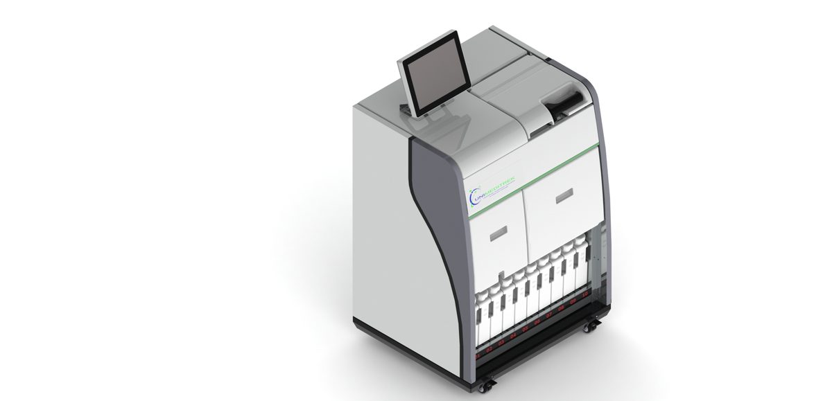
Tissue processors are essential for automating the fixation, dehydration, and clearing steps. These machines can handle multiple samples simultaneously, ensuring consistent processing conditions for each tissue. Modern tissue processors offer programmable protocols, allowing for customization based on tissue type and size. Regular maintenance and calibration of tissue processors are crucial for optimal performance.
Microtomes
Microtomes are precision instruments used for sectioning embedded tissues. There are several types of microtomes, including rotary, sliding, and ultramicrotomes, each suited for different applications. The choice of microtome depends on the desired section thickness and the type of tissue being processed. High-quality microtome blades and proper technique are essential for obtaining thin, uniform sections. Staining Machines
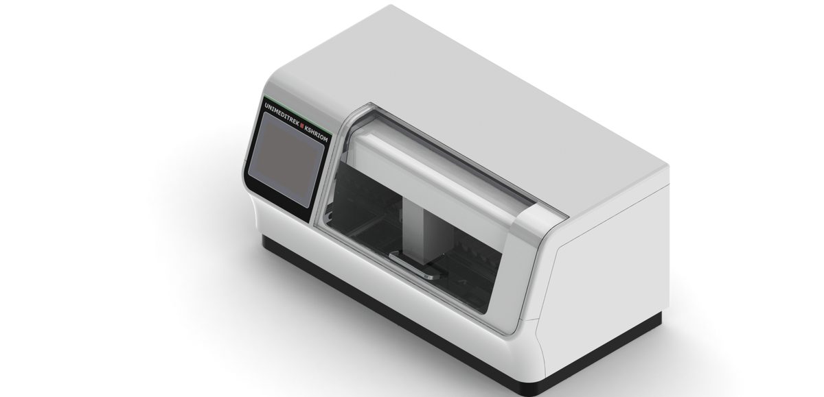
Staining machines automate the application of dyes to tissue sections, ensuring consistent staining quality. These machines can perform a variety of staining protocols, including H&E, IHC, and special stains. Automated staining machines reduce variability and improve reproducibility, making them valuable tools in high-throughput laboratories. Regular cleaning and maintenance of staining machines are necessary to prevent cross-contamination and ensure reliable results.
Conclusion and Future Trends in Tissue Processing
Mastering tissue processing is a journey that combines technical skill, attention to detail, and a deep understanding of histological principles. By following the essential steps outlined in this article, histologists can achieve high-quality tissue sections that are crucial for accurate diagnosis and research. From fixation to staining, each step in the tissue processing workflow plays a vital role in preserving cellular architecture and enhancing visibility under the microscope. Looking forward, the field of tissue processing is poised for significant advancements. Automation and digital pathology are set to revolutionize the way tissues are processed and analyzed, improving efficiency and diagnostic accuracy. Additionally, emerging staining techniques and molecular methods will expand the capabilities of histology, offering new insights into cellular function and disease mechanisms. Staying abreast of these innovations and incorporating them into routine practice will be essential for maintaining excellence in tissue processing. In conclusion, mastering tissue processing is not just about following protocols; it is about understanding the underlying principles and continuously striving for improvement. By embracing best practices, troubleshooting common issues, and adopting new technologies, histologists can ensure that their work contributes to the advancement of medical science and the delivery of high-quality patient care. The future of tissue processing is bright, and those who master its essential steps will be at the forefront of this exciting field.
FAQs
What are the main steps of tissue processing?
The core steps are fixation, dehydration, clearing, paraffin infiltration and embedding — done in sequence to preserve tissue morphology and produce a block that sections cleanly.
Why is tissue processing important in histopathology?
It preserves cellular architecture so thin, well-stained, artefact-free sections can be examined under the microscope — block quality directly affects diagnostic accuracy.
Prepared and reviewed by the Unimeditrek technical team based on laboratory practice, installation and service experience. This document explains histopathology workflow, laboratory practice and equipment based on the questions we hear from hospitals, medical colleges and diagnostic laboratories — vendor-neutral where it explains the science and practical where it explains equipment.
Prepared by Unimeditrek Pvt. Ltd.. For product specifications and quotations, contact our team.