Home › Knowledge › Mastering Correct Tissue Orientation at Embedding
Knowledge articleMastering Correct Tissue Orientation at Embedding
Essential techniques for histopathology labs
 Tissue Embedding Station — Unimeditrek" style="max-width:100%;border-radius:10px"/>
Introduction to Tissue Orientation
Tissue orientation is a critical step in the histopathology workflow, particularly during the embedding phase. Proper orientation ensures that the cellular architecture of the tissue is preserved, allowing for accurate diagnosis and analysis. The goal is to position the tissue in such a way that the most relevant areas are available for sectioning, which directly impacts the quality of the histological slides produced.
When tissues are embedded, the way they are oriented can affect the visibility of structures within the sample. For instance, in a biopsy of a tumor, the orientation can determine whether the pathologist can see the invasive edges of the tumor or the relationship of the tumor to surrounding tissues. Understanding the principles of correct tissue orientation can significantly enhance the diagnostic yield of histopathological examinations.
Techniques for Achieving Proper Orientation
To achieve proper tissue orientation during embedding, several techniques can be employed. One of the most common methods is to use the "face" of the tissue, which is the cut surface that best represents the tissue's architecture. This surface should be placed facing the embedding mold. For larger specimens, it may be necessary to trim the tissue to expose the most informative areas before embedding.
Another important consideration is the use of orientation markers. These can be small pieces of tissue or specific inks that help the technician identify the correct orientation during the cutting process. Markers can be particularly useful in complex specimens where multiple layers or types of tissue are present. By ensuring that the orientation is clear, pathologists can avoid unnecessary confusion during the sectioning process.
Common Mistakes in Tissue Orientation
Despite the best intentions, mistakes in tissue orientation can occur. One common error is embedding the tissue in a way that does not allow for the optimal view of the area of interest. For example, embedding a tumor sample upside down can lead to missing critical details that are vital for diagnosis.
Another frequent mistake is not considering the natural contours of the tissue. Tissues often have irregular shapes, and forcing them into a mold without accounting for their natural form can lead to distortion. This distortion can compromise the integrity of the sections and result in a loss of valuable diagnostic information. Recognizing these potential pitfalls is essential for improving the embedding process and ensuring high-quality histological slides.
Best Practices for Embedding
Implementing best practices can greatly enhance the quality of tissue embedding. One key practice is to ensure that the embedding molds are appropriately sized for the specimens. Using molds that are too large can lead to poor support for the tissue, while molds that are too small can cause compression and distortion.
Additionally, maintaining a clean and organized workspace is crucial. Contaminants can affect the embedding medium and, ultimately, the quality of the tissue sections. Regularly cleaning the embedding station and ensuring that all tools are well-maintained can help mitigate these issues. Furthermore, it is advisable to practice embedding with a variety of tissue types to develop a keen sense of how best to orient different specimens.
Conclusion and Practical Takeaway
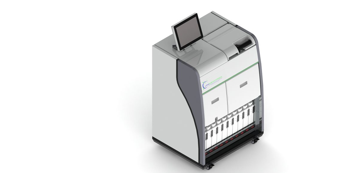
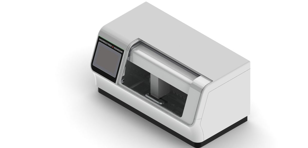
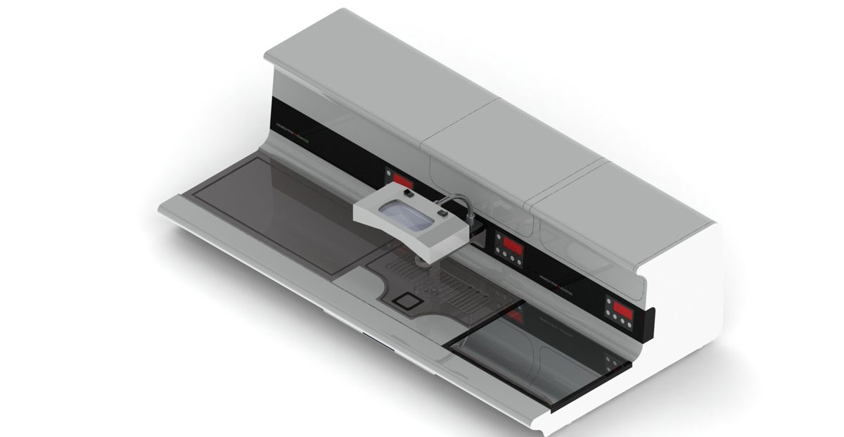
Correct tissue orientation at embedding is a vital aspect of histopathological practice. By employing proper techniques, avoiding common mistakes, and adhering to best practices, histopathology labs can ensure that their diagnostic processes are as accurate and efficient as possible. The use of a tissue embedding station, such as those supplied by Unimeditrek, can aid in achieving optimal orientation, ultimately leading to better patient outcomes.
As a practical takeaway, always take the time to assess your tissue specimens before embedding. Identifying the most informative areas and using orientation markers can save time and enhance the quality of your histological sections. Remember, the goal is to present the tissue in a way that allows for the best possible evaluation by the pathologist.
FAQs
What is the importance of tissue orientation during embedding?
Tissue orientation is crucial as it ensures that the most relevant areas of the sample are available for sectioning, which impacts the accuracy of diagnosis.
How can I avoid mistakes in tissue orientation?
To avoid mistakes, carefully assess the tissue morphology, use orientation markers, and ensure the embedding mold is appropriate for the specimen size.
What are some best practices for embedding tissue samples?
Best practices include maintaining a clean workspace, using appropriately sized molds, and practicing with various tissue types to enhance your embedding skills.
Prepared and reviewed by the Unimeditrek technical team based on laboratory practice, installation and service experience. This document explains histopathology workflow, laboratory practice and equipment based on the questions we hear from hospitals, medical colleges and diagnostic laboratories — vendor-neutral where it explains the science and practical where it explains equipment.
Prepared by Unimeditrek Pvt. Ltd.. For product specifications and quotations, contact our team.