Home › Knowledge › Introduction to IHC Controls and Reproducibility
Knowledge articleIntroduction to IHC Controls and Reproducibility
Understanding the Essentials for Effective Immunohistochemistry
Understanding Immunohistochemistry (IHC)
Immunohistochemistry (IHC) is a powerful technique utilized in histopathology to detect specific antigens in tissue sections. This method relies on the use of antibodies that bind to target proteins, allowing for the visualization of cellular components. The accuracy of IHC results is heavily dependent on the quality of the controls used during the staining process. Understanding the role of these controls is crucial for pathologists and laboratory technicians alike, as they help ensure the reliability and reproducibility of the results.
In IHC, controls are typically classified into positive and negative controls. Positive controls are samples known to express the target antigen, confirming that the staining protocol is functioning correctly. Conversely, negative controls are samples that do not express the target antigen, ensuring that any observed staining is specific and not due to non-specific binding of the antibody. The selection of appropriate controls is essential for validating the staining process and interpreting results accurately.
The Importance of Reproducibility
Reproducibility in IHC is vital for maintaining the integrity of diagnostic results. It refers to the ability to obtain consistent results across multiple tests and conditions. Factors influencing reproducibility include the quality of reagents, the precision of the staining protocol, and the skill of the personnel conducting the tests. A lack of reproducibility can lead to misdiagnosis and affect patient management, making it imperative for laboratories to establish standardized protocols and practices.
To enhance reproducibility, laboratories should implement robust quality control measures. This includes regular calibration and maintenance of equipment, such as the digital tissue floatation bath, which is crucial for preparing tissue sections. Consistency in sample handling, fixation methods, and antigen retrieval techniques also plays a significant role in achieving reproducible results. By focusing on these elements, labs can improve the reliability of their IHC assays.
Implementing Effective IHC Controls
When implementing IHC controls, it is important to select appropriate tissue samples that reflect the range of antigen expression expected in clinical specimens. For positive controls, tissues known to express the target antigen, such as tumor tissues or specific organ tissues, should be used. This ensures that the assay is capable of detecting the antigen under optimal conditions. Negative controls, on the other hand, should ideally include tissues that are known not to express the target antigen to rule out any false positives.
Moreover, documenting the performance of controls is essential for ongoing quality assurance. Laboratories should maintain records of control results alongside patient samples to track trends and identify any deviations in assay performance. This documentation can help in troubleshooting issues and refining protocols over time, ultimately leading to improved diagnostic accuracy.
The Role of Equipment in IHC
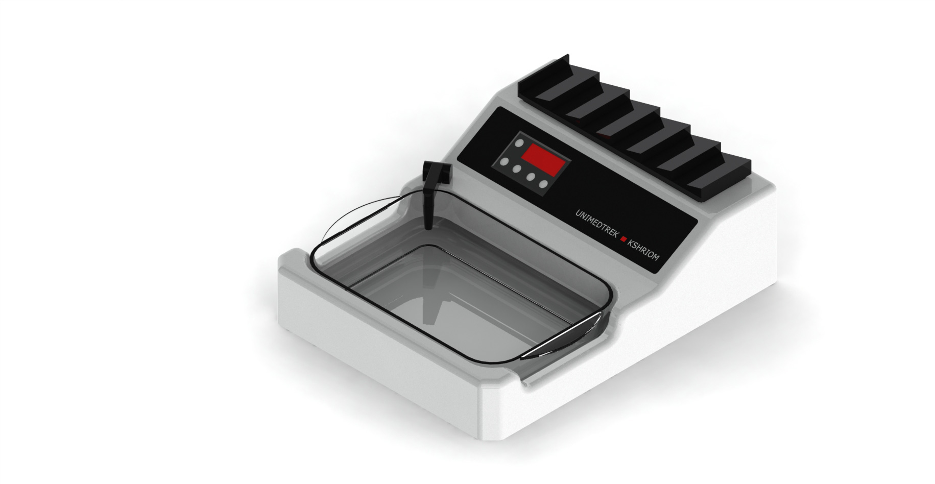
The equipment used in IHC, including a digital tissue floatation bath, plays a significant role in the preparation of tissue sections. The floatation bath is designed to facilitate the smooth handling of sections and ensure that they are adequately spread out on slides. Proper sectioning and mounting are critical for achieving optimal staining results, as uneven sections can lead to inconsistent staining patterns.
In addition to sectioning, the temperature control provided by the digital tissue floatation bath is crucial for maintaining the integrity of the tissue samples. Maintaining an appropriate temperature prevents the degradation of antigens and ensures that they remain accessible for antibody binding. Regular maintenance and calibration of the floatation bath are essential to ensure its reliability in the staining process.
Practical Takeaway for Laboratories
For laboratories aiming to improve their IHC practices, focusing on controls and reproducibility is paramount. Establishing a routine for quality control, including the use of positive and negative controls, will help validate the staining process and ensure accurate results. Additionally, investing in reliable equipment, such as a digital tissue floatation bath, can enhance the consistency of tissue section preparation.
Moreover, continuous education and training for laboratory personnel are essential for maintaining high standards in IHC practices. Encouraging a culture of quality and accountability will help laboratories achieve better outcomes in patient diagnostics and research applications.
Further reading
For peer-reviewed depth on histopathology practice and quality, these leading journals are excellent references:
- The Journal of Pathology
- Histopathology (Wiley)
- Modern Pathology (USCAP)
- Archives of Pathology & Laboratory Medicine (CAP)
- American Journal of Clinical Pathology
- Journal of Clinical Pathology (BMJ)
Tissue Floatation Bath — engineered and serviced in India
Unimeditrek manufactures the Tissue Floatation Bath with pan-India installation and service. Explore the full specifications, request a demo, or register to download the brochure — direct from the manufacturer.
View Tissue Floatation Bath & request a quote →FAQs
What are positive and negative controls in IHC?
Positive controls are samples known to express the target antigen, confirming that the staining protocol works. Negative controls are samples that do not express the target antigen, ensuring observed staining is specific.
How can I improve reproducibility in my IHC assays?
Improving reproducibility involves standardizing protocols, maintaining equipment, and ensuring consistent sample handling, fixation, and antigen retrieval techniques.
Why is the digital tissue floatation bath important for IHC?
The digital tissue floatation bath is crucial for preparing tissue sections, ensuring they are evenly spread on slides, which is vital for achieving optimal staining results.
Prepared and reviewed by the Unimeditrek technical team based on laboratory practice, installation and service experience. This document explains histopathology workflow, laboratory practice and equipment based on the questions we hear from hospitals, medical colleges and diagnostic laboratories — vendor-neutral where it explains the science and practical where it explains equipment.
Prepared by Unimeditrek Pvt. Ltd.. For product specifications and quotations, contact our team.