Home › Knowledge › Formalin Fixation: Timing, Volume, and Penetration Insights
Knowledge articleFormalin Fixation: Timing, Volume, and Penetration Insights
Essential tips for effective tissue preparation
Understanding Formalin Fixation
Formalin fixation is a critical step in the histopathology workflow, serving to preserve tissue morphology and prevent autolysis. The process involves immersing tissue samples in a formaldehyde solution, which cross-links proteins and stabilizes cellular structures. This fixation is essential for accurate diagnosis, as it ensures that the tissue's architecture and cellular details remain intact for subsequent examination under a microscope.
However, achieving optimal fixation requires careful attention to several factors, including time, volume of formalin, and penetration. Each of these elements plays a significant role in the quality of the fixed tissue, impacting the staining characteristics and overall interpretability of histological slides. Understanding the nuances of formalin fixation can greatly enhance the effectiveness of the tissue floatation bath used in preparing samples for microscopy.
Time: The Crucial Factor
The duration of fixation is paramount; under-fixation can lead to poor preservation, while over-fixation might mask cellular details. Generally, the fixation time should be proportional to the size of the tissue specimen. Smaller biopsies may require only a few hours, while larger specimens can take longer. A common guideline is to allow a minimum of 6 to 8 hours for adequate fixation of standard-sized tissues, but this can vary based on the specific type of tissue and its density.
It is also essential to monitor the fixation process closely. For instance, if tissues are fixed for too long, they may become overly hard and brittle, complicating sectioning and staining. This is where the tissue floatation bath comes into play, as it aids in the gentle manipulation of fixed tissues, allowing for optimal sectioning and preservation of delicate structures. Regularly checking the status of tissue samples during fixation can help ensure that they receive the appropriate duration of exposure to formalin.
Volume: Ensuring Adequate Fixative
The volume of formalin used in the fixation process is another critical aspect that can influence the quality of tissue preservation. A general rule of thumb is to use a volume of fixative that is at least 10 to 20 times greater than the volume of the tissue specimen. This ensures that the formalin can adequately permeate the tissue and facilitate effective fixation throughout the entire specimen.
Inadequate volume can lead to uneven fixation, resulting in areas of necrosis or autolysis within the same specimen. This is particularly important when dealing with larger or denser tissues, which may require more formalin to achieve thorough penetration. Lab technicians should be mindful of this when preparing samples, as the use of insufficient fixative can lead to compromised tissue quality, ultimately affecting diagnostic outcomes.
Penetration: Achieving Uniform Fixation
Effective penetration of formalin into the tissue is essential for uniform fixation. Factors such as tissue thickness, density, and the type of formalin solution can affect how well the fixative penetrates. Thicker tissues may require additional time and volume for complete fixation, as the formalin needs to diffuse through the tissue layers.
To enhance penetration, some laboratories use techniques such as cutting larger specimens into smaller pieces or utilizing a more concentrated formalin solution. Additionally, ensuring that the tissue is fully submerged in the fixative and agitating the container can help promote even distribution of the formalin. Proper fixation is crucial for preserving the integrity of the cellular architecture, which is vital for accurate histological assessment.
Integration with the Tissue Floatation Bath
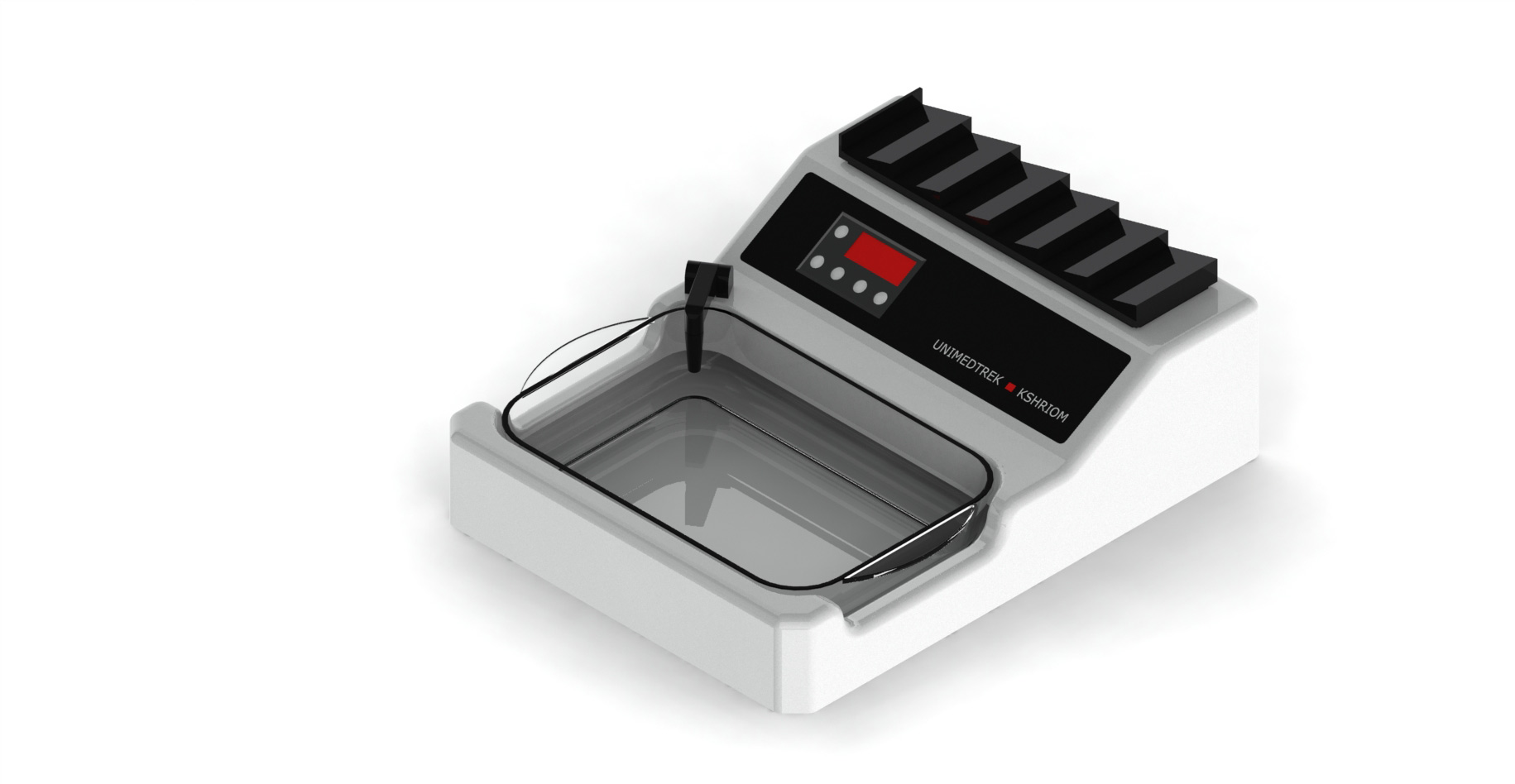
The tissue floatation bath is an invaluable tool in the histopathology lab, especially after formalin fixation. After fixation, tissues are often dehydrated and embedded in paraffin wax, and the tissue floatation bath allows for the gentle manipulation of tissue sections, facilitating even spreading and preventing distortion. This step is critical for obtaining high-quality sections that are suitable for staining and microscopic evaluation.
When using the tissue floatation bath, it is essential to maintain the correct temperature to ensure optimal tissue handling. The bath should be set to a temperature that allows for the melting of paraffin without overheating the tissue, which can lead to loss of morphological details. Proper technique in using the tissue floatation bath can significantly enhance the quality of histological slides and contribute to more accurate diagnostic results.
Practical Takeaway
In conclusion, mastering the nuances of formalin fixation—time, volume, and penetration—is essential for histopathology labs aiming to produce high-quality tissue samples. Keeping these factors in mind, along with the effective use of the tissue floatation bath, can lead to improved outcomes in tissue preparation and ultimately enhance diagnostic accuracy.
Further reading
For peer-reviewed depth on histopathology practice and quality, these leading journals are excellent references:
- The Journal of Pathology
- Histopathology (Wiley)
- Modern Pathology (USCAP)
- Archives of Pathology & Laboratory Medicine (CAP)
- American Journal of Clinical Pathology
- Journal of Clinical Pathology (BMJ)
Tissue Floatation Bath — engineered and serviced in India
Unimeditrek manufactures the Tissue Floatation Bath with pan-India installation and service. Explore the full specifications, request a demo, or register to download the brochure — direct from the manufacturer.
View Tissue Floatation Bath & request a quote →FAQs
What is the ideal fixation time for tissue specimens?
The ideal fixation time varies by tissue size, but generally, smaller specimens require around 6 to 8 hours, while larger ones may need longer.
How much formalin should I use for fixation?
Use a volume of formalin that is at least 10 to 20 times greater than the tissue volume to ensure adequate penetration and fixation.
How does the tissue floatation bath assist in histopathology?
The tissue floatation bath aids in the gentle manipulation of fixed tissue sections, ensuring even spreading and preventing distortion during the embedding process.
Prepared and reviewed by the Unimeditrek technical team based on laboratory practice, installation and service experience. This document explains histopathology workflow, laboratory practice and equipment based on the questions we hear from hospitals, medical colleges and diagnostic laboratories — vendor-neutral where it explains the science and practical where it explains equipment.
Prepared by Unimeditrek Pvt. Ltd.. For product specifications and quotations, contact our team.