Home › Knowledge › Formalin Fixation: Optimizing Time, Volume, and Penetration
Knowledge articleFormalin Fixation: Optimizing Time, Volume, and Penetration
A guide for effective tissue embedding practices
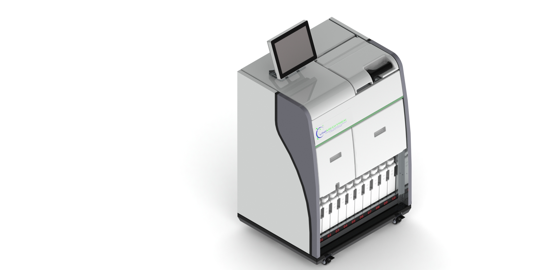
 Tissue Embedding Station — Unimeditrek" style="max-width:100%;border-radius:10px"/>
Understanding Formalin Fixation
Formalin fixation is a critical step in the histopathology process that preserves tissue morphology and cellular details for microscopic examination. It involves immersing tissue samples in formaldehyde solutions, primarily to prevent autolysis and putrefaction. The fixation process stabilizes proteins and cross-links them, creating a network that retains the tissue's structural integrity. Without proper fixation, the subsequent analyses may yield inaccurate or inconclusive results, making it essential for pathologists and lab technicians to understand the nuances of this process.
The effectiveness of formalin fixation depends on several factors, including the time of fixation, the volume of formalin used, and the penetration of the fixative into the tissue. Each of these elements plays a significant role in ensuring that the tissue is adequately preserved for embedding and sectioning. The optimal combination of these factors can vary based on the type and size of the tissue sample, as well as the specific requirements of the downstream histological techniques.
Time of Fixation
The duration of fixation is crucial, as under-fixing can lead to poor preservation of cellular details, while over-fixing may cause tissue hardening, making it difficult to cut thin sections. Generally, the recommended fixation time can vary based on the size and type of tissue. Smaller specimens may require only a few hours, while larger specimens might need overnight fixation. It is essential to monitor the fixation process closely to avoid these common pitfalls.
In many labs, a standard protocol is established to guide technicians on the appropriate fixation times for various tissue types. However, it is also important to consider the specific needs of each sample. For example, delicate tissues may require shorter fixation times to prevent the development of artifacts. Pathologists should always be aware that the fixation time is not a one-size-fits-all approach and should adjust protocols based on their observations and results.
Volume of Formalin
The volume of formalin used during fixation is another critical factor. A common guideline is to use a volume of formalin that is at least 10 to 20 times greater than the volume of the tissue specimen. This ensures that the fixative can penetrate adequately and uniformly throughout the tissue. Inadequate volume can lead to uneven fixation, which can compromise the quality of the histological sections.
Moreover, using too much formalin can lead to wastage and increased costs. Pathologists and lab technicians should strive to find a balance between sufficient volume for effective fixation and minimizing waste. It is also advisable to consider the container's size and shape, as these factors can affect the distribution of formalin around the tissue. Ensuring that the tissue is fully immersed in formalin will enhance fixation efficiency.
Penetration of Fixative
The penetration of formalin into the tissue is vital for achieving optimal fixation. The rate of penetration can be influenced by several factors, including the thickness of the tissue, the concentration of formalin, and the temperature at which fixation occurs. Thicker tissues may take longer to fix due to the time required for the formalin to diffuse through the sample. As a general rule, thinner sections will fix more rapidly than thicker ones.
To improve penetration, it can be beneficial to cut larger specimens into smaller pieces that are more manageable. Additionally, using a higher concentration of formalin can enhance penetration but may also increase the risk of over-fixing. Therefore, it is essential to strike a balance and monitor the fixation process closely. Some labs may also employ techniques such as agitation or vacuum infiltration to facilitate better penetration of the fixative.
Integrating Fixation with the Tissue Embedding Station
Once the fixation process is complete, the tissue is ready for the next steps in histopathology, which typically include dehydration, clearing, and embedding. A tissue embedding station plays a crucial role in this phase, as it provides the necessary environment for embedding tissues in paraffin wax. Properly fixed tissues will yield better results during embedding, ensuring that the cellular architecture is preserved for sectioning.
When using a tissue embedding station, it is essential to ensure that the samples are adequately dehydrated and that the embedding medium is of high quality. This will facilitate the cutting of thin sections that are vital for accurate diagnosis. Additionally, technicians should follow the established protocols for embedding to maintain consistency and quality across samples. Proper integration of the fixation process with the embedding station will ultimately lead to improved histopathological outcomes.
Practical Takeaway
Optimizing formalin fixation requires careful consideration of time, volume, and penetration. By adhering to best practices and adjusting protocols based on specific tissue types and sizes, pathologists and lab technicians can enhance the quality of their histopathological analyses. A well-executed fixation process lays the foundation for successful embedding and accurate diagnosis.
Further reading
For peer-reviewed depth on histopathology practice and quality, these leading journals are excellent references:
- The Journal of Pathology
- Histopathology (Wiley)
- Modern Pathology (USCAP)
- Archives of Pathology & Laboratory Medicine (CAP)
- American Journal of Clinical Pathology
- Journal of Clinical Pathology (BMJ)
Tissue Embedding Station — engineered and serviced in India
Unimeditrek manufactures the Tissue Embedding Station with pan-India installation and service. Explore the full specifications, request a demo, or register to download the brochure — direct from the manufacturer.
View Tissue Embedding Station & request a quote →FAQs
What is the ideal fixation time for different tissue types?
The ideal fixation time varies based on the tissue type and size. Smaller samples may need a few hours, while larger samples could require overnight fixation.
How can I ensure proper penetration of formalin in larger specimens?
To ensure proper penetration, consider cutting larger specimens into smaller pieces and using a sufficient volume of formalin to fully immerse the tissue.
What role does the tissue embedding station play in histopathology?
The tissue embedding station is essential for embedding fixed tissues in paraffin, ensuring that cellular architecture is preserved for accurate sectioning.
Prepared and reviewed by the Unimeditrek technical team based on laboratory practice, installation and service experience. This document explains histopathology workflow, laboratory practice and equipment based on the questions we hear from hospitals, medical colleges and diagnostic laboratories — vendor-neutral where it explains the science and practical where it explains equipment.
Prepared by Unimeditrek Pvt. Ltd.. For product specifications and quotations, contact our team.