Home › Knowledge › Formalin Fixation: Mastering Time, Volume, and Penetration
Knowledge articleFormalin Fixation: Mastering Time, Volume, and Penetration
Essential tips for optimal histology results
 Slide Warming Table — Unimeditrek" style="max-width:100%;border-radius:10px"/>
Understanding Formalin Fixation
Formalin fixation is a crucial step in the histopathology workflow, serving to preserve tissue specimens for microscopic examination. The primary purpose of formalin is to cross-link proteins, thereby stabilizing the cellular structure and preventing autolysis and putrefaction. This preservation is essential for obtaining accurate diagnoses, as it maintains the morphology of tissues and cells, allowing pathologists to observe pathological changes with precision.
To achieve optimal fixation, it’s important to consider the type of formalin used, typically a 10% neutral buffered formalin (NBF), which has a pH of around 7.0. This buffering helps to prevent the formation of formaldehyde precipitates that can interfere with tissue processing. Additionally, the choice of formalin concentration can affect the quality of fixation, as higher concentrations may lead to tissue shrinkage and hardening, while lower concentrations may not adequately preserve the tissue.
Time: Finding the Right Balance
The fixation time is a critical factor that can significantly influence the quality of tissue preservation. If tissue specimens are left in formalin for too short a duration, they may not be adequately fixed, leading to poor morphology and compromised staining characteristics. Conversely, excessive fixation can result in over-hardening of the tissue, making it difficult to cut thin sections during the embedding process.
As a general guideline, the fixation time should be proportional to the size of the tissue specimen. Smaller biopsies may require only a few hours of fixation, while larger specimens can take 24 hours or more. It is essential to monitor the fixation process and ensure that specimens are not left in formalin longer than necessary. Utilizing a histology hot plate can aid in the fixation process by maintaining a consistent temperature, which can enhance the penetration of formalin into the tissue.
Volume: The Importance of Adequate Formalin
Another critical aspect of formalin fixation is the volume of formalin used relative to the size of the tissue specimen. A common recommendation is to use at least 10 times the volume of formalin compared to the specimen size. This ensures that formalin can adequately penetrate the tissue and create a uniform fixation. Insufficient formalin volume can lead to uneven fixation, resulting in artifacts and diagnostic challenges.
When preparing specimens, it’s also important to ensure that the formalin is well-mixed and free of debris. Any sediment or particulate matter can interfere with the fixation process and may cause inconsistencies in tissue preservation. Using a dedicated histology hot plate can facilitate this process by providing a stable environment for mixing and maintaining the formalin at an optimal temperature for penetration.
Pentration: Ensuring Effective Tissue Fixation
The penetration of formalin into the tissue is a vital component of effective fixation. Factors such as tissue thickness, density, and even the method of specimen handling can influence how well formalin penetrates. Thicker tissues may require additional time and volume for proper fixation, as formalin must diffuse into the tissue to achieve effective cross-linking of proteins.
To enhance penetration, consider cutting larger specimens into smaller pieces before fixation. This increases the surface area exposed to formalin and allows for more rapid and uniform penetration. Additionally, using a histology hot plate during the fixation process can help maintain a consistent temperature that promotes better formalin diffusion into the tissue.
Practical Takeaway: Optimizing Your Fixation Protocol
In summary, mastering the nuances of formalin fixation is essential for achieving high-quality histological specimens. By paying close attention to the time, volume, and penetration of formalin, histopathology labs can significantly improve their diagnostic accuracy. Implementing the use of a histology hot plate can further enhance the fixation process, ensuring that tissue specimens are optimally preserved for microscopic analysis.
Always remember to evaluate each specimen individually, as variations in tissue type and size may necessitate adjustments to your fixation protocol. Regularly reviewing and refining your fixation practices can lead to improved outcomes and more reliable histopathological diagnoses.
Further reading
For peer-reviewed depth on histopathology practice and quality, these leading journals are excellent references:
- The Journal of Pathology
- Histopathology (Wiley)
- Modern Pathology (USCAP)
- Archives of Pathology & Laboratory Medicine (CAP)
- American Journal of Clinical Pathology
- Journal of Clinical Pathology (BMJ)
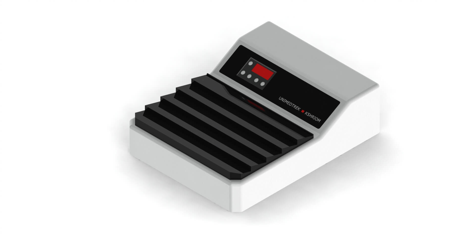
Slide Warming Table — engineered and serviced in India
Unimeditrek manufactures the Slide Warming Table with pan-India installation and service. Explore the full specifications, request a demo, or register to download the brochure — direct from the manufacturer.
View Slide Warming Table & request a quote →FAQs
What is the ideal fixation time for tissue specimens?
The ideal fixation time varies based on the size and type of tissue, but generally, smaller specimens require a few hours while larger ones may need 24 hours or more.
How does the volume of formalin affect fixation quality?
Using at least 10 times the volume of formalin compared to the tissue size ensures adequate penetration and uniform fixation, preventing artifacts.
Why is penetration important in formalin fixation?
Proper penetration ensures that formalin reaches all parts of the tissue, allowing for effective cross-linking of proteins and preserving cellular morphology.
Prepared and reviewed by the Unimeditrek technical team based on laboratory practice, installation and service experience. This document explains histopathology workflow, laboratory practice and equipment based on the questions we hear from hospitals, medical colleges and diagnostic laboratories — vendor-neutral where it explains the science and practical where it explains equipment.
Prepared by Unimeditrek Pvt. Ltd.. For product specifications and quotations, contact our team.