Home › Knowledge › Decalcification of Bone and Calcified Tissue: A Primer
Knowledge articleDecalcification of Bone and Calcified Tissue: A Primer
Essential techniques for histopathology labs
 Automatic Slide Staining Machine — Unimeditrek" style="max-width:100%;border-radius:10px"/>
Understanding Decalcification
Decalcification is a critical process in histopathology, particularly when dealing with bone and calcified tissues. The primary objective of decalcification is to remove calcium deposits from tissues, allowing for easier sectioning and improved staining quality. This process is crucial for obtaining clear, high-quality histological slides that reveal the underlying cellular architecture. Without proper decalcification, the presence of calcium can interfere with the penetration of stains, leading to suboptimal results.
Decalcification can be achieved through various methods, including chemical, physical, and enzymatic techniques. Chemical decalcification typically involves the use of acids, such as hydrochloric acid or nitric acid, which dissolve calcium salts. Physical methods include the use of ultrasonic waves or microwave energy to facilitate the decalcification process. Each method has its advantages and limitations, and the choice often depends on the specific requirements of the tissue sample and the downstream applications.
Choosing the Right Decalcification Agent
The selection of an appropriate decalcifying agent is paramount to the success of the decalcification process. Common acids used for decalcification include formic acid, ethylenediaminetetraacetic acid (EDTA), and citric acid. Each of these agents has distinct properties that can influence the rate of decalcification and the quality of the final histological preparation. For instance, formic acid is effective for rapid decalcification but may cause tissue shrinkage, while EDTA is gentler and preserves tissue morphology better.
It is also essential to consider the concentration of the decalcifying agent and the duration of exposure. Overexposure to strong acids can lead to tissue damage, while underexposure may result in incomplete decalcification. Monitoring the decalcification process is crucial; many labs employ a technique called "poking" to assess the progress of decalcification, which involves using a needle to check the hardness of the bone.
Optimizing the Decalcification Process
To achieve optimal results during decalcification, it is essential to establish a standard protocol tailored to the specific types of tissues being processed. Factors such as the size of the specimen, the type of decalcifying agent, and the desired speed of decalcification should all be taken into account. A common practice is to start with a lower concentration of acid and gradually increase it as needed, ensuring that tissue integrity is maintained throughout the process.
Additionally, the temperature at which decalcification occurs can impact the efficiency of the process. Warmer temperatures can accelerate the action of the decalcifying agent, but care must be taken to avoid excessive heat that could damage the tissue. Regularly checking the pH of the decalcifying solution can also help ensure that the conditions remain optimal for effective decalcification.
Preparing for Slide Staining
Once decalcification is complete, the next step is preparing the tissue for embedding and sectioning. It is vital to wash the decalcified tissue thoroughly to remove any residual acid, as leftover acid can adversely affect the staining process. Typically, tissues are rinsed in buffered saline or distilled water, followed by dehydration through graded alcohols, which prepares the tissue for embedding in paraffin.
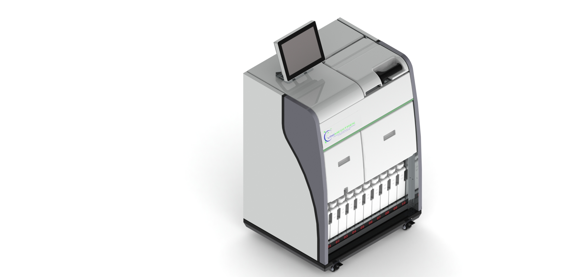
The use of an automatic slide staining machine can significantly enhance the efficiency and consistency of the staining process. These machines provide precise control over staining times and temperatures, which are critical for achieving optimal results. Moreover, they reduce the variability associated with manual staining, ensuring that each slide is processed uniformly, which is particularly important when comparing results across multiple samples.
Practical Takeaway
In summary, effective decalcification is essential for producing high-quality histological slides from bone and calcified tissues. By understanding the various methods and agents available, pathologists and lab technicians can optimize the decalcification process to preserve tissue integrity and improve staining outcomes. Implementing standardized protocols and utilizing tools like an automatic slide staining machine can further enhance the reliability and efficiency of histopathological analyses.
Further reading
For peer-reviewed depth on histopathology practice and quality, these leading journals are excellent references:
- The Journal of Pathology
- Histopathology (Wiley)
- Modern Pathology (USCAP)
- Archives of Pathology & Laboratory Medicine (CAP)
- American Journal of Clinical Pathology
- Journal of Clinical Pathology (BMJ)
Automatic Slide Staining Machine — engineered and serviced in India
Unimeditrek manufactures the Automatic Slide Staining Machine with pan-India installation and service. Explore the full specifications, request a demo, or register to download the brochure — direct from the manufacturer.
View Automatic Slide Staining Machine & request a quote →FAQs
What is the best method for decalcifying bone tissue?
The best method for decalcifying bone tissue depends on the specific requirements of the histological analysis, with common techniques including chemical decalcification using acids or EDTA.
How do I know when decalcification is complete?
Decalcification can be assessed by checking the hardness of the tissue; a needle can be used to poke the specimen, and if it feels soft, decalcification is likely complete.
Can I use an automatic slide staining machine for decalcified tissues?
Yes, an automatic slide staining machine is well-suited for decalcified tissues, as it ensures consistent staining results and reduces variability in the staining process.
Prepared and reviewed by the Unimeditrek technical team based on laboratory practice, installation and service experience. This document explains histopathology workflow, laboratory practice and equipment based on the questions we hear from hospitals, medical colleges and diagnostic laboratories — vendor-neutral where it explains the science and practical where it explains equipment.
Prepared by Unimeditrek Pvt. Ltd.. For product specifications and quotations, contact our team.