Home › Knowledge › Decalcification of Bone and Calcified Tissue: A Practical Primer
Knowledge articleDecalcification of Bone and Calcified Tissue: A Practical Primer
Essential techniques for effective histopathology
 Slide Warming Table — Unimeditrek" style="max-width:100%;border-radius:10px"/>
Introduction to Decalcification
Decalcification is a critical process in histopathology, particularly when dealing with bone and calcified tissues. The primary goal of decalcification is to remove calcium deposits from tissues, allowing for better infiltration of embedding media and clearer visualization of cellular structures under a microscope. This process is essential when preparing samples for histological examination, as the presence of calcium can obscure tissue architecture and hinder the accuracy of diagnoses.
In histopathology labs, the choice of decalcifying agent and method can significantly affect the quality of the final slides. Various agents, such as acids and chelating agents, are commonly used, each with unique properties and effects on the tissue. Understanding the mechanisms of these agents is crucial for pathologists and lab technicians to ensure optimal results in their work.
Common Decalcifying Agents
There are several decalcifying agents available, with the most widely used being hydrochloric acid and nitric acid. Hydrochloric acid is effective and relatively quick, making it a popular choice in many laboratories. However, it can lead to tissue damage if not monitored closely, as it may affect cellular morphology. Nitric acid, while also effective, tends to produce more artifacts and can cause excessive tissue shrinkage.
Chelating agents like EDTA (ethylenediaminetetraacetic acid) are another option, offering a gentler alternative to acid-based decalcifiers. EDTA works by binding calcium ions, effectively removing them without the harsh effects of acids. This method is particularly advantageous for preserving the integrity of delicate tissues and cellular structures, making it a preferred choice for many histopathologists.
Decalcification Techniques
Choosing the right technique for decalcification is essential for achieving high-quality histological slides. The most common methods include immersion, continuous flow, and microwave-assisted decalcification. Immersion involves placing the tissue in a decalcifying solution for a specified period, which can vary based on the size and density of the specimen.
Continuous flow decalcification utilizes a constant flow of fresh decalcifying solution over the tissue, which can expedite the process and enhance the quality of the results. Microwave-assisted decalcification is a newer technique that employs microwave energy to accelerate the decalcification process, significantly reducing the time required. Each method has its advantages and limitations, and the choice often depends on the specific requirements of the tissue being processed.
Importance of Monitoring Decalcification
Monitoring the decalcification process is crucial to ensure that the tissue is adequately decalcified without overexposure to the decalcifying agent. Over-decalcification can lead to tissue damage, loss of cellular detail, and ultimately, compromised diagnostic accuracy. It is essential to check the tissue regularly during the process, using techniques such as X-ray or chemical tests to determine the level of calcium removal.
Once the tissue has reached the desired level of decalcification, it should be thoroughly rinsed to remove any residual decalcifying agent. This step is vital to prevent interference with subsequent staining procedures, which can be negatively impacted by leftover chemicals. Proper handling and monitoring throughout the process contribute to the overall quality of the histological slides produced.
Utilizing the Slide Warming Table in Decalcification
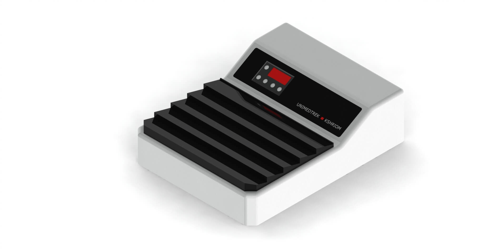
The slide warming table can play a significant role in the histopathological process, particularly in the context of decalcification. By providing a controlled environment for warming slides, it aids in the optimal drying and adherence of tissue sections to slides after decalcification and embedding. Proper warming can enhance the quality of the staining process, leading to clearer and more detailed histological evaluations.
Furthermore, maintaining an appropriate temperature during the preparation of slides can prevent issues such as condensation and moisture retention, which can adversely affect tissue quality. Utilizing a slide warming table ensures that the slides are adequately prepared for microscopic examination, ultimately improving diagnostic outcomes.
Practical Takeaway
Decalcification is a vital step in the histopathology workflow, particularly for bone and calcified tissues. Understanding the different decalcifying agents, techniques, and the importance of monitoring the process can significantly enhance the quality of histological slides. Utilizing tools like the slide warming table can further improve the preparation of these slides, ensuring that pathologists have the best possible samples for accurate diagnoses.
Further reading
For peer-reviewed depth on histopathology practice and quality, these leading journals are excellent references:
- The Journal of Pathology
- Histopathology (Wiley)
- Modern Pathology (USCAP)
- Archives of Pathology & Laboratory Medicine (CAP)
- American Journal of Clinical Pathology
- Journal of Clinical Pathology (BMJ)
Slide Warming Table — engineered and serviced in India
Unimeditrek manufactures the Slide Warming Table with pan-India installation and service. Explore the full specifications, request a demo, or register to download the brochure — direct from the manufacturer.
View Slide Warming Table & request a quote →FAQs
What is the best decalcifying agent for bone tissue?
The best decalcifying agent depends on the specific requirements of the tissue and the desired outcomes. Hydrochloric acid is commonly used for its effectiveness, but EDTA is preferred for preserving delicate structures.
How long should I decalcify bone specimens?
The duration of decalcification varies based on the size and density of the specimen. Regular monitoring is essential to achieve optimal results without over-decalcifying.
Can I use a microwave for decalcification?
Yes, microwave-assisted decalcification is a viable technique that can significantly reduce the time required for decalcification while maintaining tissue integrity.
Prepared and reviewed by the Unimeditrek technical team based on laboratory practice, installation and service experience. This document explains histopathology workflow, laboratory practice and equipment based on the questions we hear from hospitals, medical colleges and diagnostic laboratories — vendor-neutral where it explains the science and practical where it explains equipment.
Prepared by Unimeditrek Pvt. Ltd.. For product specifications and quotations, contact our team.