Home › Knowledge › Decalcification of Bone and Calcified Tissue: A Practical Primer
Knowledge articleDecalcification of Bone and Calcified Tissue: A Practical Primer
Essential techniques for histopathology labs
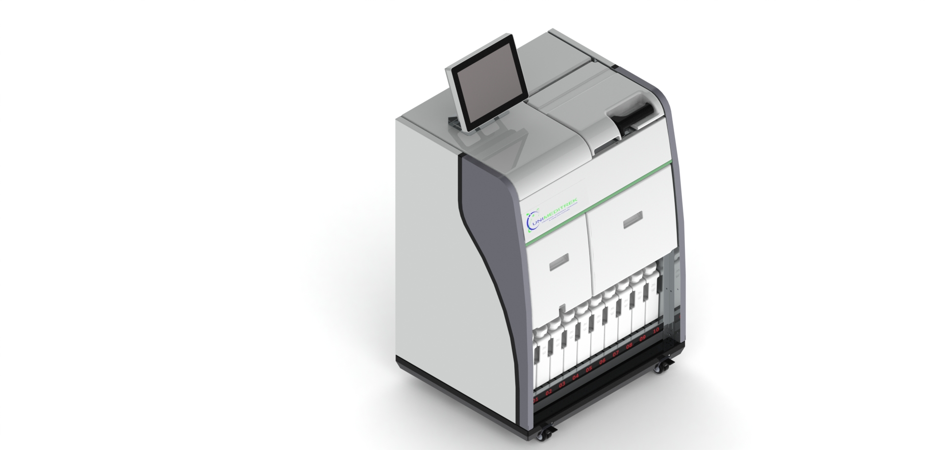
 Tissue Embedding Station — Unimeditrek" style="max-width:100%;border-radius:10px"/>
Introduction to Decalcification
Decalcification is a crucial step in histopathology, especially when preparing bone and calcified tissues for microscopic examination. The primary goal of decalcification is to remove calcium deposits while preserving the overall morphology of the tissue. This process is vital because calcified tissues can obscure cellular details, making accurate diagnosis challenging. Understanding the principles of decalcification and the various methods available is essential for pathologists and lab technicians alike.
In histopathology, the decalcification process can be influenced by several factors, including the type of tissue, the fixation method used, and the subsequent embedding techniques. Proper decalcification ensures that tissues can be adequately infiltrated with embedding media, such as paraffin, allowing for optimal sectioning and staining. This primer aims to provide practical insights into effective decalcification strategies and their integration with the use of a tissue embedder.
Methods of Decalcification
There are several methods for decalcifying bone and calcified tissues, each with its advantages and disadvantages. The two primary categories of decalcification methods are chemical and physical. Chemical decalcification typically involves the use of acids, such as hydrochloric acid or nitric acid, which dissolve calcium salts. These acids can be used in various concentrations and can be adjusted based on the specific tissue type and desired outcome.
Physical decalcification methods, on the other hand, include techniques such as ion exchange and ultrasonic waves. Ion exchange methods involve the use of chelating agents that bind to calcium ions, effectively removing them from the tissue. Ultrasonic decalcification utilizes high-frequency sound waves to enhance the penetration of decalcifying agents, resulting in faster decalcification times. Understanding the nuances of these methods allows histopathology professionals to choose the most appropriate technique for their specific needs.
Factors Influencing Decalcification
Several factors can significantly influence the decalcification process, including tissue thickness, type of fixative used, and the duration of exposure to decalcifying agents. Thicker specimens may require longer decalcification times, while the choice of fixative can impact the ease of decalcification. For instance, tissues fixed in formalin may decalcify more slowly than those fixed in alcohol-based solutions.
Moreover, the temperature at which decalcification occurs can also play a role. Warmer temperatures can accelerate the decalcification process, but care must be taken to avoid tissue damage or alteration. Regular monitoring of the tissue during decalcification is essential to ensure that the desired level of decalcification is achieved without compromising tissue integrity. By understanding these factors, lab technicians can optimize their decalcification protocols to achieve the best results.
Embedding Decalcified Tissues
Once decalcification is complete, the next step is embedding the tissue in a suitable medium. The use of a tissue embedder is critical in this stage, as it ensures that the decalcified tissue is properly infiltrated with embedding media, such as paraffin. Proper embedding is essential for achieving thin, uniform sections that are suitable for microscopic examination.
It is important to note that decalcified tissues may require specific embedding protocols to accommodate their altered physical properties. For example, decalcified bone may be more fragile than non-decalcified bone, necessitating careful handling during the embedding process. Additionally, the choice of embedding medium can affect the quality of the final sections, making it essential to select a medium that provides optimal support for the decalcified tissue.
Practical Takeaway
In summary, effective decalcification of bone and calcified tissues is a critical component of histopathological practice. Understanding the various methods available, the factors influencing decalcification, and the importance of proper embedding techniques can significantly enhance the quality of tissue samples for microscopic analysis. By integrating these practices into daily workflows, histopathology professionals can ensure accurate diagnoses and improve patient outcomes.
Further reading
For peer-reviewed depth on histopathology practice and quality, these leading journals are excellent references:
- The Journal of Pathology
- Histopathology (Wiley)
- Modern Pathology (USCAP)
- Archives of Pathology & Laboratory Medicine (CAP)
- American Journal of Clinical Pathology
- Journal of Clinical Pathology (BMJ)
Tissue Embedding Station — engineered and serviced in India
Unimeditrek manufactures the Tissue Embedding Station with pan-India installation and service. Explore the full specifications, request a demo, or register to download the brochure — direct from the manufacturer.
View Tissue Embedding Station & request a quote →FAQs
What is the best method for decalcifying bone tissue?
The best method for decalcifying bone tissue depends on various factors, including the type of tissue and the desired outcome. Chemical methods using acids are commonly used, but physical methods like ion exchange can also be effective.
How long does the decalcification process typically take?
The duration of the decalcification process can vary widely based on the method used, tissue thickness, and type of fixative. Regular monitoring is essential to achieve the desired level of decalcification without compromising tissue integrity.
Can decalcified tissues be embedded in paraffin?
Yes, decalcified tissues can be embedded in paraffin, but it is important to ensure that they are adequately infiltrated with the embedding medium to achieve high-quality sections suitable for microscopic examination.
Prepared and reviewed by the Unimeditrek technical team based on laboratory practice, installation and service experience. This document explains histopathology workflow, laboratory practice and equipment based on the questions we hear from hospitals, medical colleges and diagnostic laboratories — vendor-neutral where it explains the science and practical where it explains equipment.
Prepared by Unimeditrek Pvt. Ltd.. For product specifications and quotations, contact our team.