Home › Knowledge › Cryostat Microtome: Daily Use in Histopathology Labs
Use caseCryostat Microtome: Daily Use in Histopathology Labs
A vital tool for precise tissue analysis
Introduction to Cryostat Microtome
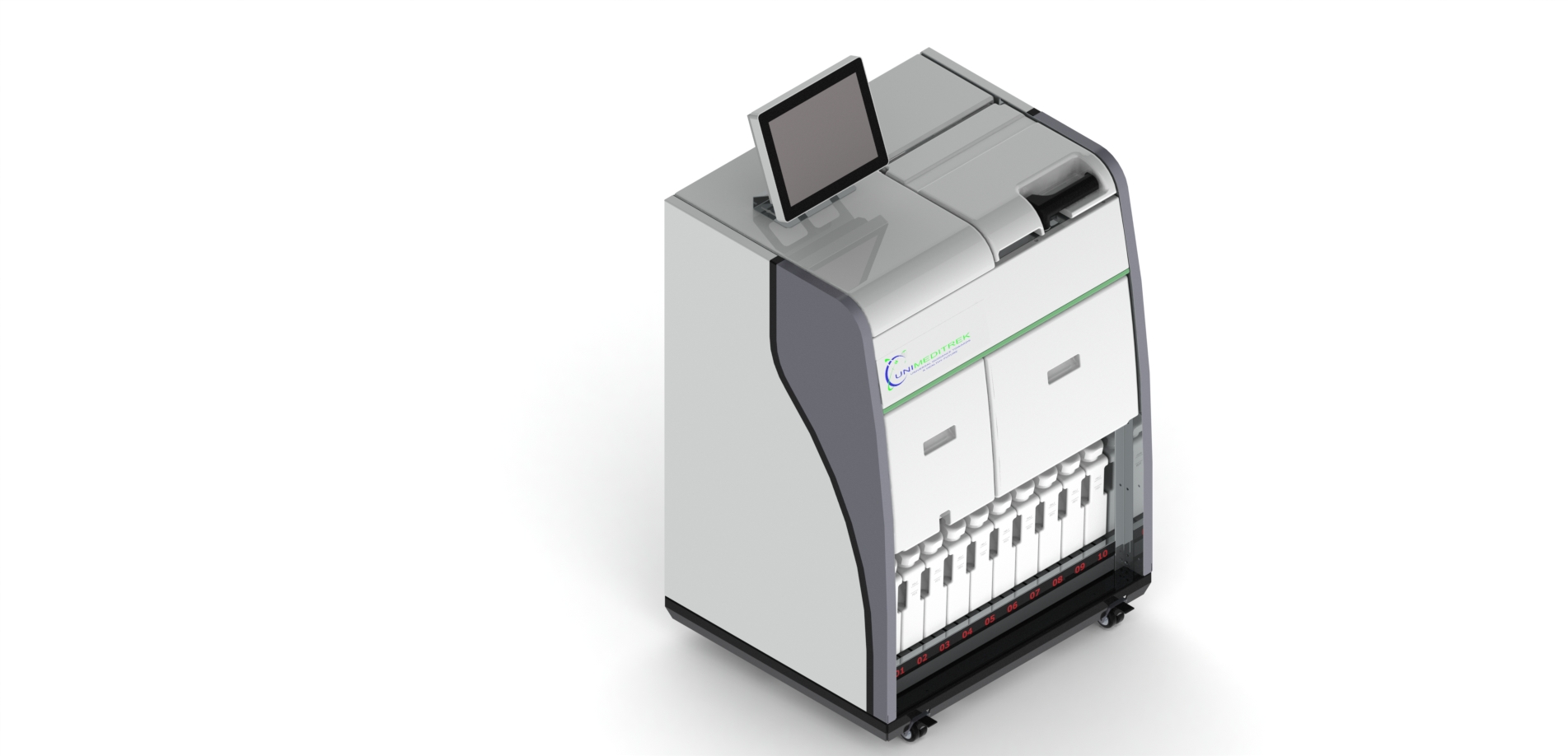
The Cryostat Microtome is an essential piece of equipment in histopathology labs, particularly in India where rapid diagnosis is crucial for effective patient care. This device allows pathologists to prepare thin sections of frozen tissue for microscopic examination, ensuring that accurate diagnoses can be made swiftly.
Daily Workflow in a Histopathology Lab
In a typical histopathology lab, the Cryostat Microtome plays a pivotal role in the daily workflow. After a surgical procedure, tissue samples are often sent for immediate analysis. The Cryostat Microtome is utilized to freeze the tissue samples quickly, allowing for the preparation of sections that can be examined under a microscope within hours.
Step-by-Step Process
- Tissue Preparation: The tissue sample is first embedded in a cryoprotectant medium to preserve its cellular integrity.
- Freezing: The sample is then placed in the Cryostat Microtome, where it is rapidly frozen to maintain its structural details.
- Sectioning: Once frozen, the microtome’s blade is adjusted to cut ultra-thin sections, typically between 5 to 10 micrometers thick.
- Mounting: The sections are then mounted on glass slides for staining and further analysis.
Advantages of Using Cryostat Microtome
The Cryostat Microtome offers several advantages that significantly enhance the efficiency of histopathology labs. Firstly, the ability to prepare frozen sections allows for immediate examination, which is critical in intraoperative consultations. This speed can be vital in guiding surgical decisions.
Additionally, the precision of the Cryostat Microtome ensures that the tissue sections maintain their morphological characteristics, which is essential for accurate diagnosis. The ease of operation and maintenance also makes it a preferred choice among laboratory technicians.
Challenges and Considerations
While the Cryostat Microtome is an invaluable tool, it does come with its challenges. Maintaining the optimal temperature is crucial to avoid ice crystal formation, which can compromise tissue quality. Regular calibration and maintenance are necessary to ensure the microtome operates effectively and produces consistent results.
Moreover, technicians must be trained adequately to handle the device, as improper usage can lead to damaged samples or inaccurate sectioning.
Conclusion and Practical Takeaway
The Cryostat Microtome is a cornerstone of modern histopathology, facilitating rapid and accurate tissue analysis in Indian labs. By integrating this technology into daily workflows, pathologists can significantly improve diagnostic turnaround times, ultimately enhancing patient care.
Practical Takeaway: For histopathology labs looking to enhance their efficiency, investing in a high-quality Cryostat Microtome and ensuring proper training for staff can lead to improved diagnostic outcomes and patient satisfaction.
FAQs
What is a Cryostat Microtome used for?
A Cryostat Microtome is used to cut thin sections of frozen tissue for histopathological examination.
How does the Cryostat Microtome improve diagnostic speed?
It allows for rapid preparation of tissue samples, enabling pathologists to provide quicker diagnoses during surgical procedures.
Prepared and reviewed by the Unimeditrek technical team based on laboratory practice, installation and service experience. This document explains histopathology workflow, laboratory practice and equipment based on the questions we hear from hospitals, medical colleges and diagnostic laboratories — vendor-neutral where it explains the science and practical where it explains equipment.
Prepared by Unimeditrek Pvt. Ltd.. For product specifications and quotations, contact our team.