Home › Knowledge › Common Tissue Processing Artefacts and Their Causes
Knowledge articleCommon Tissue Processing Artefacts and Their Causes
Understanding issues with your automatic tissue processor
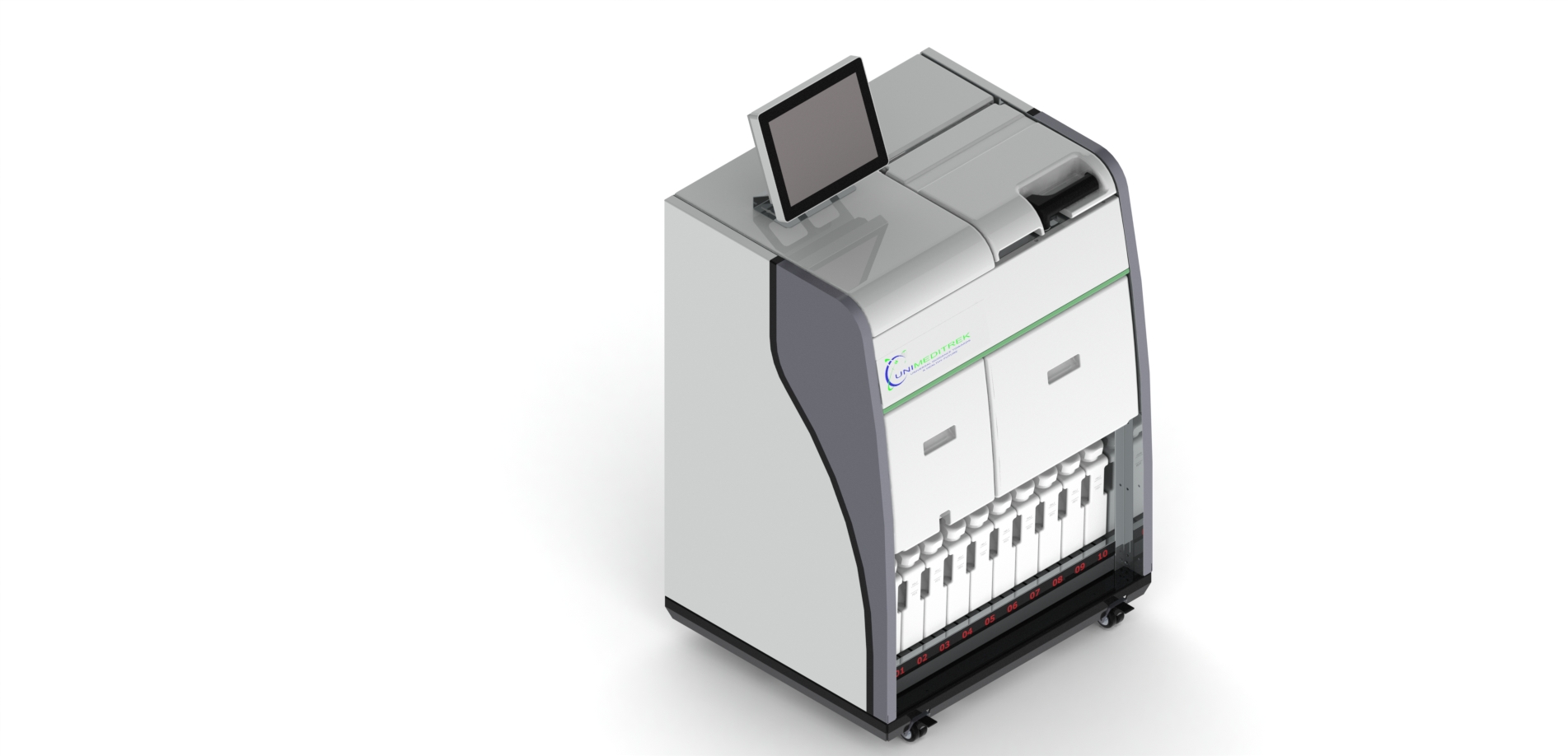
Introduction to Tissue Processing Artefacts
Tissue processing is a critical step in histopathology that involves preparing biological specimens for microscopic examination. During this process, artefacts can occur, which may compromise the quality of the tissue sections and lead to misinterpretations. Understanding these artefacts, their causes, and how to mitigate them is essential for histopathologists and lab technicians working with an automatic tissue processor.
Artefacts can arise from various stages of tissue processing, including fixation, dehydration, clearing, infiltration, and embedding. Each stage presents unique challenges that can lead to artefacts if not managed properly. In this article, we will explore some of the most common artefacts encountered in histopathology, their underlying causes, and practical solutions to prevent them.
Common Artefacts in Tissue Processing
One of the most frequently observed artefacts is the "shrinkage" of tissue specimens. This occurs when the tissue loses water during the dehydration phase, leading to a reduction in size. Shrinkage can result in loss of morphological details, making it difficult to accurately assess the tissue under a microscope. Factors contributing to shrinkage include excessive dehydration time, high concentration of dehydrating agents, or inadequate fixation prior to processing.
Another common artefact is "folding" or "crumpling" of tissue sections. This can occur during the embedding process, particularly if the embedding medium is not adequately infiltrated or if the tissue is not properly oriented. Folding can obscure important cellular details and hinder diagnosis. To avoid this, ensure that the embedding medium is heated appropriately and that the tissue is placed in the correct orientation during embedding.
Causes of Artefacts in Tissue Processing
Artefacts can also arise from improper fixation, which is a crucial step in preserving tissue morphology. Inadequate fixation can lead to "autolysis," where enzymes within the tissue begin to break down cellular structures before processing is complete. This breakdown can create artefacts that mimic pathological changes, leading to potential misdiagnosis. To prevent autolysis, it is essential to use an appropriate fixative and to ensure that fixation occurs promptly after tissue collection.
Additionally, temperature fluctuations during processing can contribute to artefacts. If the automatic tissue processor operates outside the recommended temperature range, it may affect the chemical reactions necessary for proper tissue preservation. For instance, if the clearing agent is not adequately heated, it may not fully infiltrate the tissue, leading to incomplete clearing and artefacts in the final sections. Regular calibration and maintenance of the automatic tissue processor can help mitigate these risks.
Impact of Artefacts on Diagnosis
The presence of artefacts can significantly impact the diagnostic accuracy of histopathological evaluations. Artefacts may mimic or obscure pathological features, leading to misinterpretation of the tissue's condition. For example, shrinkage artefacts may cause a pathologist to misjudge the extent of a lesion, while folding artefacts can hide critical cellular details necessary for an accurate diagnosis.
Moreover, artefacts can lead to unnecessary additional testing, increased workload, and ultimately, delays in patient care. Therefore, it is crucial for histopathologists and lab technicians to be aware of these artefacts and their causes to ensure high-quality tissue processing. Regular training and quality control measures can help minimize the occurrence of artefacts and improve diagnostic outcomes.
Practical Solutions to Minimize Artefacts
To minimize artefacts during tissue processing, it is essential to establish standard operating procedures (SOPs) that include best practices for each stage of the process. This includes using appropriate fixatives, maintaining consistent processing times, and ensuring that the automatic tissue processor is regularly serviced and calibrated. Training staff on the importance of each step in the processing workflow can also enhance the overall quality of tissue preparation.
Additionally, implementing quality control measures, such as routine checks for artefacts in processed tissue sections, can help identify issues early in the process. By fostering a culture of quality assurance within the lab, histopathologists and technicians can work together to minimize artefacts and enhance the accuracy of their diagnostic work.
Practical Takeaway
Understanding common tissue processing artefacts and their causes is crucial for ensuring the quality of histopathological evaluations. By implementing best practices and regularly maintaining the automatic tissue processor, labs can significantly reduce the occurrence of artefacts and improve diagnostic accuracy.
Further reading
For peer-reviewed depth on histopathology practice and quality, these leading journals are excellent references:
- The Journal of Pathology
- Histopathology (Wiley)
- Modern Pathology (USCAP)
- Archives of Pathology & Laboratory Medicine (CAP)
- American Journal of Clinical Pathology
- Journal of Clinical Pathology (BMJ)
Automatic Tissue Processor — engineered and serviced in India
Unimeditrek manufactures the Automatic Tissue Processor with pan-India installation and service. Explore the full specifications, request a demo, or register to download the brochure — direct from the manufacturer.
View Automatic Tissue Processor & request a quote →FAQs
What are the most common artefacts seen in tissue processing?
Common artefacts include shrinkage, folding, and autolysis of tissue specimens, which can affect the quality of histopathological evaluations.
How can I prevent artefacts during tissue processing?
To prevent artefacts, ensure proper fixation, maintain consistent processing times, and regularly calibrate the automatic tissue processor.
What impact do artefacts have on histopathological diagnosis?
Artefacts can obscure critical cellular details and mimic pathological changes, potentially leading to misinterpretation and misdiagnosis.
Prepared and reviewed by the Unimeditrek technical team based on laboratory practice, installation and service experience. This document explains histopathology workflow, laboratory practice and equipment based on the questions we hear from hospitals, medical colleges and diagnostic laboratories — vendor-neutral where it explains the science and practical where it explains equipment.
Prepared by Unimeditrek Pvt. Ltd.. For product specifications and quotations, contact our team.


