Home › Knowledge › Common H&E and Section Artefacts: Causes and Fixes
Knowledge articleCommon H&E and Section Artefacts: Causes and Fixes
Understanding Issues in Histopathology
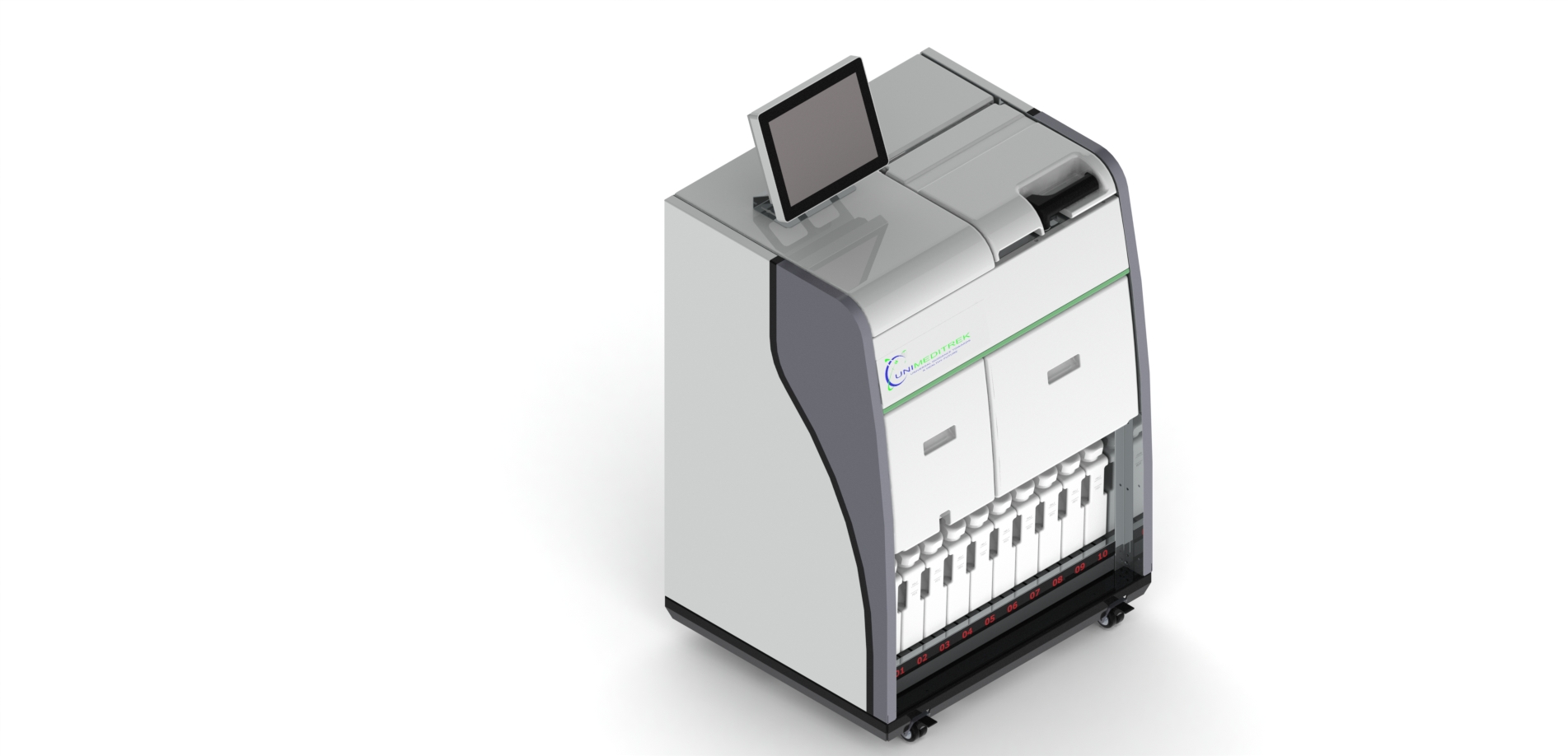
 Tissue Embedding Station — Unimeditrek" style="max-width:100%;border-radius:10px"/>
Introduction to H&E Staining and Sectioning
Hematoxylin and eosin (H&E) staining is a cornerstone of histopathology, providing critical insights into tissue morphology. However, the process is not without its challenges, often leading to artefacts that can obscure diagnostic clarity. Understanding these artefacts is crucial for pathologists and lab technicians alike, as it allows for improved interpretation of results and enhances the overall quality of histological evaluations.
The tissue embedding station plays a significant role in this process, aiding in the preparation of high-quality tissue sections. When artefacts arise, they can stem from various stages of tissue processing, embedding, and sectioning. Identifying the root causes of these artefacts is essential for implementing effective solutions and ensuring the integrity of histological samples.
Common Artefacts in H&E Staining
One prevalent artefact encountered during H&E staining is uneven staining, which can result from inadequate fixation or poor quality of reagents. If the tissue is not fixed properly, it may lead to differential staining, where some areas appear darker or lighter than others. This inconsistency can mislead pathologists, potentially affecting diagnosis.
To mitigate uneven staining, it is vital to ensure that tissues are adequately fixed. This involves not only the duration of fixation but also the choice of fixative. Regularly checking the quality of staining reagents and ensuring they are fresh can also help maintain consistency in results. Additionally, using a tissue embedding station that allows for uniform embedding can minimize discrepancies in tissue morphology.
Sectioning Artefacts: Causes and Solutions
Sectioning artefacts are equally problematic and can manifest in various forms, such as folds, tears, or incomplete sections. These issues often arise from improper handling of the tissue during the embedding process or incorrect settings on the microtome. For instance, using a blade that is dull can lead to uneven cuts and damage to the tissue, resulting in folds or tears.
To address these sectioning artefacts, it is crucial to maintain the microtome in optimal condition. Regularly replacing blades and ensuring they are sharp can significantly improve section quality. Moreover, adjusting the microtome settings according to the specific tissue type being sectioned is essential. For example, softer tissues may require different settings compared to harder tissues to ensure clean cuts without causing damage.
Impact of Temperature and Humidity
Environmental factors such as temperature and humidity can also contribute to artefacts in histological preparations. High humidity levels can lead to excessive water retention in tissues, causing them to swell and potentially altering their morphology. Conversely, low humidity can result in dehydration of the tissue, leading to shrinkage and distortion.
To counteract these effects, maintaining an optimal environment in the lab is paramount. This includes using climate control systems to regulate temperature and humidity levels. Additionally, ensuring that the embedding station is functioning correctly and that tissues are adequately dehydrated before embedding can help prevent artefacts related to environmental conditions.
Addressing Staining and Sectioning Artefacts
When artefacts are identified, it is essential to adopt a systematic approach to address them. This begins with a thorough review of the entire histological process, from fixation to staining and sectioning. Each stage should be scrutinized for potential sources of error, including reagent quality, fixation time, and embedding techniques.
Implementing a quality control protocol can significantly reduce the occurrence of artefacts. This may involve routinely checking the performance of the tissue embedding station and microtome, conducting regular training sessions for lab personnel, and maintaining detailed records of all procedures. By fostering a culture of quality assurance, labs can enhance the reliability of their histopathological evaluations.
Practical Takeaway
Understanding the common artefacts associated with H&E staining and sectioning is crucial for improving histopathological practices. By focusing on proper fixation, quality reagents, environmental control, and regular equipment maintenance, labs can minimize artefacts and enhance diagnostic accuracy. The tissue embedding station, when used effectively, serves as a vital tool in achieving high-quality tissue sections that are essential for reliable diagnosis.
Further reading
For peer-reviewed depth on histopathology practice and quality, these leading journals are excellent references:
- The Journal of Pathology
- Histopathology (Wiley)
- Modern Pathology (USCAP)
- Archives of Pathology & Laboratory Medicine (CAP)
- American Journal of Clinical Pathology
- Journal of Clinical Pathology (BMJ)
Tissue Embedding Station — engineered and serviced in India
Unimeditrek manufactures the Tissue Embedding Station with pan-India installation and service. Explore the full specifications, request a demo, or register to download the brochure — direct from the manufacturer.
View Tissue Embedding Station & request a quote →FAQs
What are some common artefacts seen in H&E staining?
Common artefacts in H&E staining include uneven staining, background staining, and precipitate formation, often caused by inadequate fixation or reagent quality.
How can I prevent sectioning artefacts?
To prevent sectioning artefacts, ensure the microtome blades are sharp, adjust settings according to tissue type, and handle samples gently during embedding.
What role does the tissue embedding station play in reducing artefacts?
The tissue embedding station is crucial for ensuring uniform embedding, which helps minimize artefacts during sectioning and improves overall tissue morphology.
Prepared and reviewed by the Unimeditrek technical team based on laboratory practice, installation and service experience. This document explains histopathology workflow, laboratory practice and equipment based on the questions we hear from hospitals, medical colleges and diagnostic laboratories — vendor-neutral where it explains the science and practical where it explains equipment.
Prepared by Unimeditrek Pvt. Ltd.. For product specifications and quotations, contact our team.