Home › Knowledge › Automatic Microtome: Daily Use in Histopathology Labs
Use caseAutomatic Microtome: Daily Use in Histopathology Labs
Enhancing Precision in Tissue Sectioning
Introduction to Automatic Microtome
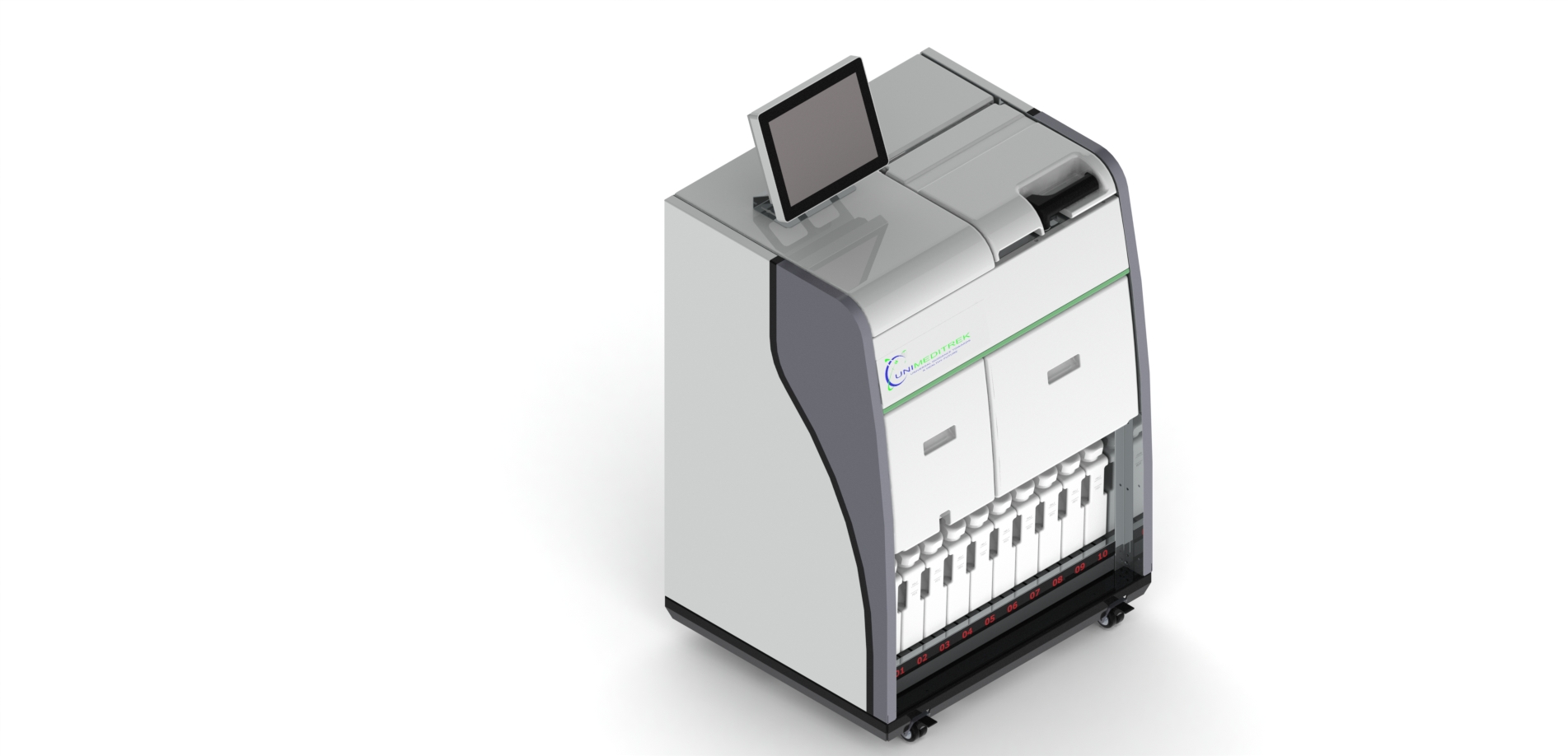
The Automatic Microtome is an essential tool in histopathology labs, designed to produce thin sections of tissue specimens for microscopic examination. Its advanced features significantly enhance the efficiency and accuracy of tissue processing, making it a staple in modern laboratories.
Daily Workflow in a Histopathology Lab
In a typical histopathology lab, the day starts with the preparation of tissue samples that have been embedded in paraffin. Once the samples are ready, the Automatic Microtome comes into play. Lab technicians set the desired thickness for the tissue sections, usually ranging from 3 to 5 micrometers, and place the paraffin block into the microtome.
The Automatic Microtome automates the cutting process, allowing for consistent and reproducible sections. This is critical as uniformity in section thickness is vital for accurate diagnosis. The use of the microtome reduces manual labor and minimizes the chances of human error, leading to higher quality samples for pathologists.
Advantages of Using Automatic Microtome
- Increased Efficiency: The Automatic Microtome can cut multiple sections in a short time, significantly speeding up the workflow.
- Precision Cuts: With programmable settings, it ensures that each section is cut to the exact specified thickness, which is crucial for histological analysis.
- Reduced Physical Strain: Automation reduces the physical demands on technicians, allowing them to focus on other critical tasks in the lab.
Real-World Application: A Day in the Lab
Consider a scenario in a busy histopathology lab in a metropolitan hospital. Each day, dozens of tissue samples arrive for analysis. The histopathology team relies on the Automatic Microtome to handle the volume efficiently. As samples are processed, technicians can quickly switch between different tissue types without the need for extensive recalibration, thanks to the microtome's user-friendly interface.
Once the sections are cut, they are placed on glass slides and stained for examination. The accuracy provided by the Automatic Microtome ensures that pathologists receive high-quality slides, which is essential for making informed diagnoses. The integration of this technology not only improves workflow but also enhances patient outcomes by facilitating timely diagnoses.
Conclusion
The Automatic Microtome has transformed the landscape of histopathology labs, providing a reliable and efficient solution for tissue sectioning. By automating the cutting process, it allows technicians to focus on analysis and interpretation, ultimately improving the quality of patient care.
Practical Takeaway
For histopathology labs looking to enhance their efficiency and accuracy, investing in an Automatic Microtome is a strategic decision that pays off in terms of quality and productivity.
FAQs
What is an Automatic Microtome used for?
An Automatic Microtome is used to cut thin sections of tissue samples for microscopic examination in histopathology.
How does an Automatic Microtome improve lab efficiency?
It automates the cutting process, allowing for faster and more consistent sectioning, which reduces manual labor and human error.
Prepared and reviewed by the Unimeditrek technical team based on laboratory practice, installation and service experience. This document explains histopathology workflow, laboratory practice and equipment based on the questions we hear from hospitals, medical colleges and diagnostic laboratories — vendor-neutral where it explains the science and practical where it explains equipment.
Prepared by Unimeditrek Pvt. Ltd.. For product specifications and quotations, contact our team.