Home › Knowledge › Artifact Identification in Histology Sections: Prevention Tips
Knowledge articleArtifact Identification in Histology Sections: Prevention Tips
Learn to identify and prevent artifacts in histology.
Understanding Artifacts in Histology Sections
Artifacts are unwanted alterations that can occur during the preparation of histology sections, potentially leading to misinterpretation of results. These artifacts can arise from various stages, including tissue fixation, processing, embedding, sectioning, and staining. Identifying these artifacts is crucial for accurate diagnosis and research outcomes.
Common Types of Artifacts
Several types of artifacts can be observed in histology sections, including:
- Compression Artifacts: Caused by excessive pressure during embedding or sectioning, leading to distorted cellular structures.
- Washing Artifacts: Resulting from inadequate washing during staining, which can cause uneven coloration.
- Air Bubbles: Formed during the embedding process, these can obscure important cellular details.
- Staining Artifacts: Occur due to improper staining techniques or reagents, leading to false positives or negatives.
Preventing Artifacts with Proper Techniques
To minimize the occurrence of artifacts, it's essential to adopt best practices throughout the histology workflow:
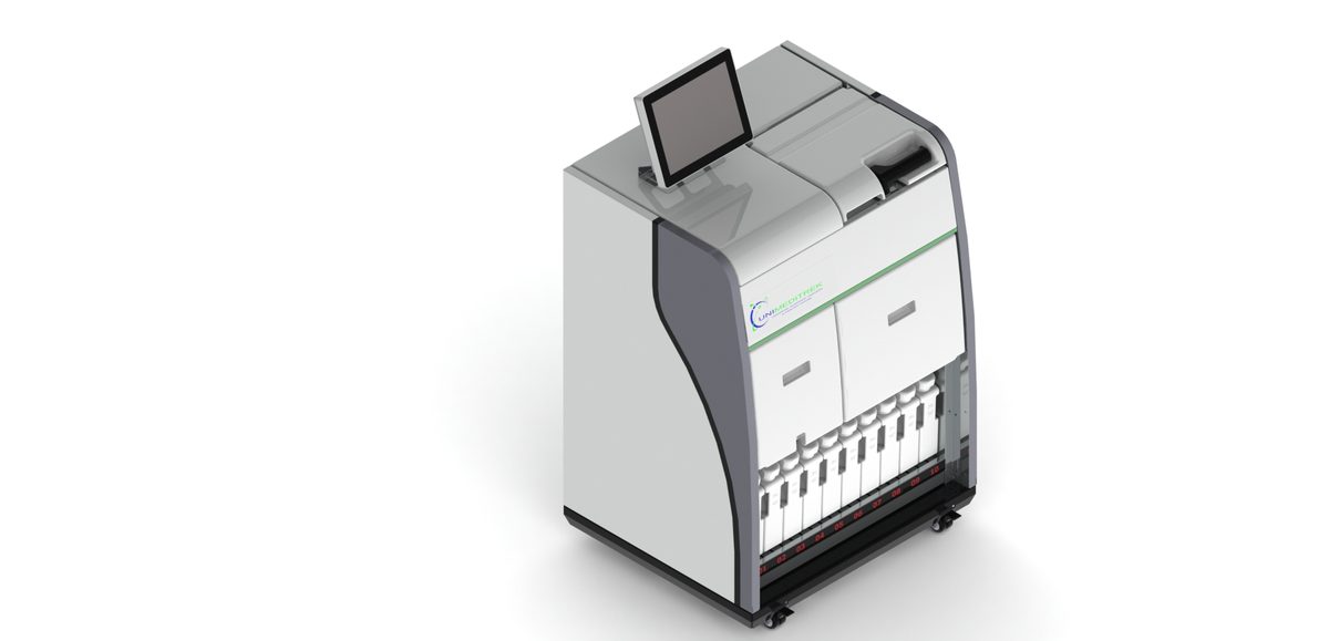
- Use an Automatic Vacuum Tissue Processor: This equipment ensures consistent fixation and dehydration, reducing the risk of compression artifacts.
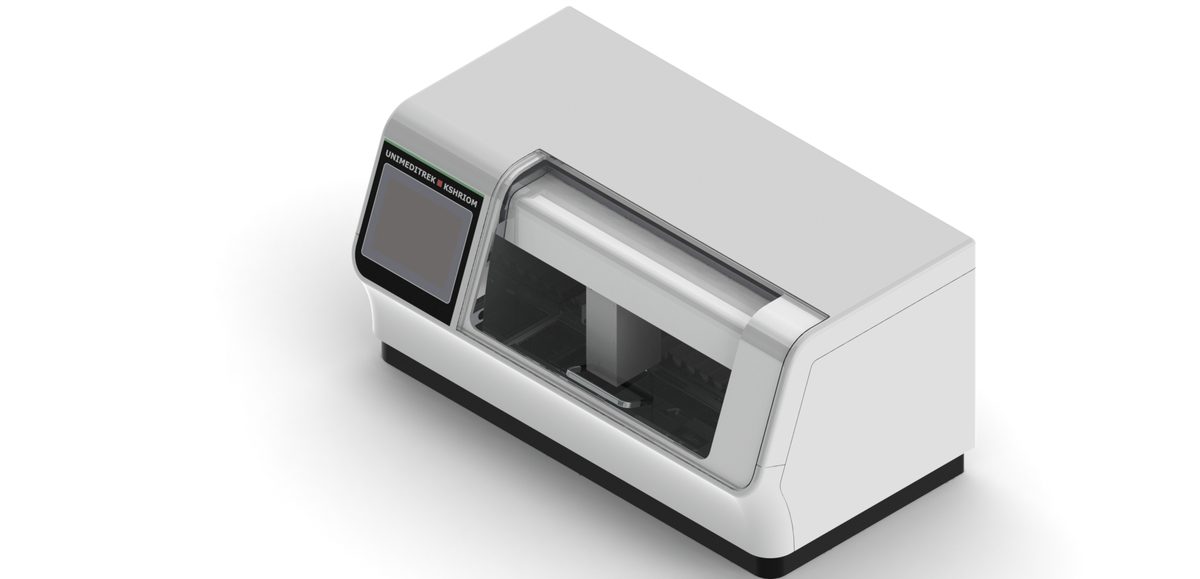
- Employ an Automatic Slide Staining Machine: Automated staining minimizes human error and provides uniform staining results, reducing staining artifacts.
- Utilize a Tissue Embedding Station: Proper embedding techniques can help prevent compression and other embedding-related artifacts.
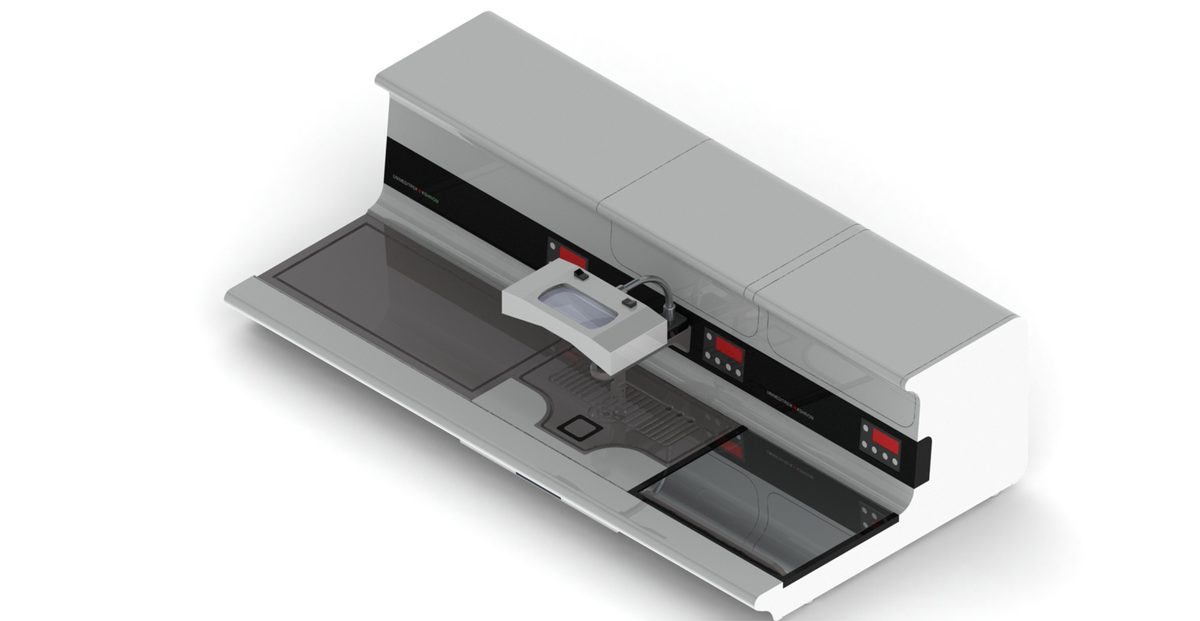
- Incorporate a Digital Tissue Floatation Bath: This helps in maintaining tissue morphology during sectioning, reducing the risk of air bubbles.
- Implement a Slide Warming Table: This aids in proper adhesion of sections to slides, preventing detachment and loss of material.
- Use a Cryostat Microtome: For precise sectioning of frozen tissues, this equipment helps maintain the integrity of delicate samples.
Identifying Artifacts During Examination
During microscopic examination, pathologists should be trained to recognize artifacts. Familiarity with the characteristics of common artifacts can enhance diagnostic accuracy. For instance, recognizing a staining artifact versus a genuine pathological change is vital for correct interpretation.
Practical Takeaway
Artifact identification and prevention are critical for achieving reliable histopathology results. By utilizing advanced equipment like the Automatic Vacuum Tissue Processor and Automatic Slide Staining Machine, histology laboratories can significantly reduce the occurrence of artifacts, ensuring high-quality diagnostic outcomes.
FAQs
What are the most common artifacts in histology?
Common artifacts include compression artifacts, washing artifacts, air bubbles, and staining artifacts.
How can I prevent artifacts in histology sections?
Use advanced equipment like Automatic Vacuum Tissue Processors and Automatic Slide Staining Machines, and follow best practices in tissue preparation.
Prepared and reviewed by the Unimeditrek technical team based on laboratory practice, installation and service experience. This document explains histopathology workflow, laboratory practice and equipment based on the questions we hear from hospitals, medical colleges and diagnostic laboratories — vendor-neutral where it explains the science and practical where it explains equipment.
Prepared by Unimeditrek Pvt. Ltd.. For product specifications and quotations, contact our team.