Standardising H&E and Routine Staining Quality
Why staining drifts, what it costs, and how automation plus reagent discipline deliver slides that look the same on every shift, operator and batch.
Abstract
Haematoxylin and eosin (H&E) is the foundation of histopathology, yet it is also one of the most variable steps in many laboratories. When staining drifts — in nuclear intensity, cytoplasmic contrast or crispness — pathologists compensate, re-read or re-stain, and teaching becomes harder. This paper explains the root causes of stain variability, the quality and cost consequences, and a practical programme of automation, reagent management and quality control that makes routine staining reproducible.
Why consistent staining matters
The pathologist reads morphology through the stain. Consistent nuclear and cytoplasmic staining is the baseline that makes subtle features reliable to interpret, makes teaching sets trustworthy, and makes quality auditable. Inconsistent staining does not just look untidy — it can mask or mimic features, and it forces avoidable re-reads and re-stains that consume reagents, slides and, most expensively, pathologist time.
Root causes of stain variability
- Operator technique: in manual staining, dip timing and agitation vary person to person and shift to shift.
- Reagent age and exhaustion: haematoxylin and differentiators change with use and time; "by-eye" reagent changes drift.
- Upstream section quality: uneven thickness, folds and poor adhesion stain unevenly however good the stainer.
- Floatation and drying: an unstable water-bath temperature or uneven drying leaves wrinkles and poor adhesion that show up as staining faults.
- Water and rinse quality: inconsistent rinsing and bluing shift the final colour balance.
A programme for reproducible staining
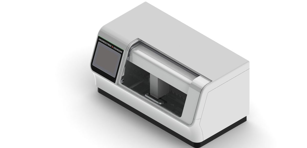
- Automate the routine. An automatic slide stainer applies identical timing, sequence and agitation to every slide, removing operator-to-operator variation and freeing technologists.
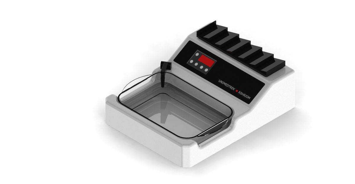
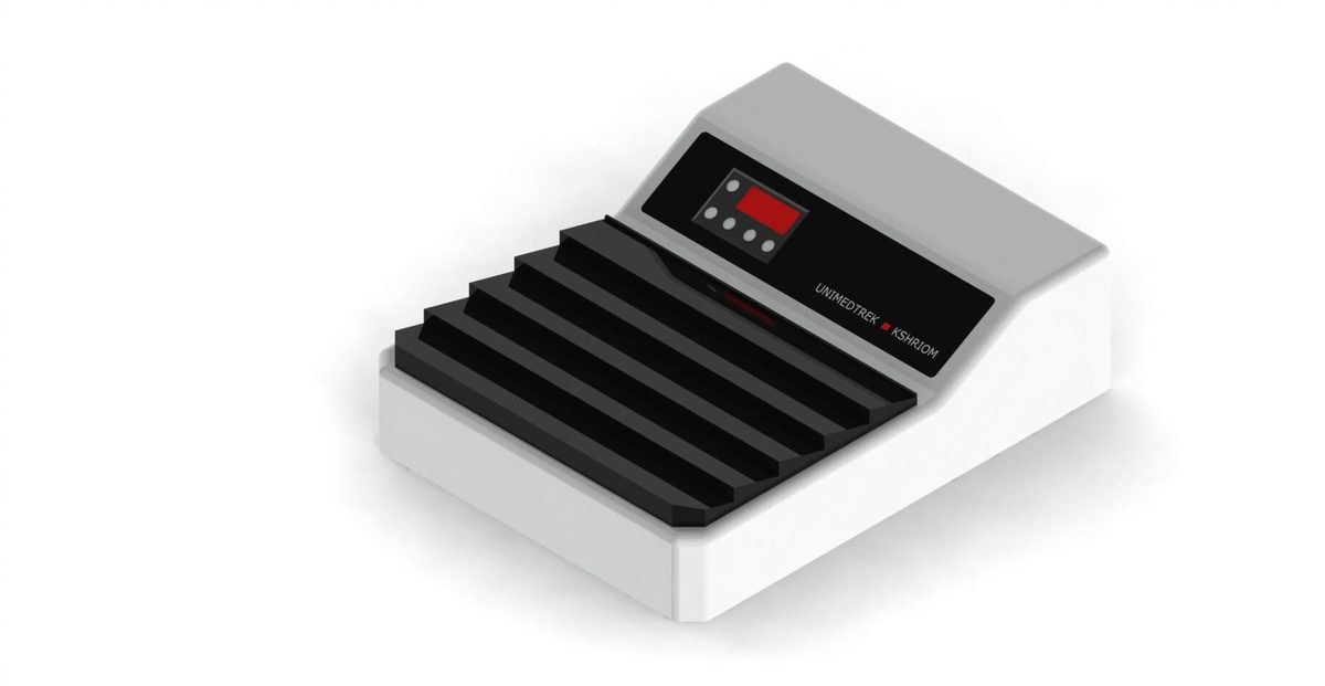
- Control the upstream steps. A temperature-stable digital floatation bath and an even warming table deliver flat, well-adhered sections that stain predictably.
- Manage reagents by schedule, not by eye. Define and document change frequencies for haematoxylin, eosin and differentiators based on slide throughput.
- Validate the protocol once, then repeat it. Establish a reference appearance and lock the protocol; change one variable at a time.
Quality control in practice
Run a known control slide periodically and keep a simple visual QC record so drift is caught early. Record reagent-change dates and lot numbers. When the appearance shifts, a documented protocol and reagent log make the cause quick to find — usually reagent exhaustion or a changed upstream step — rather than a guessing game across the whole process.
Parameters that most affect the result
Floatation water is typically held just below the paraffin melt point (around 40–45 °C); drift here is a common, under-recognised cause of "staining" faults. Sections cut evenly at the routine 3–5 µm stain far more uniformly than thick or ragged ones. And reagent change frequency, matched to throughput, is usually the dominant lever on day-to-day consistency.
How Unimeditrek helps
Our automatic slide-staining machine standardises the staining sequence itself, while our digital tissue floatation bath and slide warming table stabilise the upstream steps that quietly drive stain quality. Together with matched consumables and service support, they make reproducible H&E achievable as a routine, not an aspiration. Explore the equipment range or talk to a specialist about a staining-standardisation plan.
Frequently asked questions
Why does H&E staining drift between runs?
Mostly from operator-to-operator dip-timing variation in manual staining, reagent age and "by-eye" changes, and upstream issues — uneven section thickness, poor adhesion or an unstable floatation-bath temperature.
How does an automatic stainer improve consistency?
It applies identical timing, sequence and agitation to every slide, removing shift-to-shift variation and freeing technologists, so contrast is reproducible batch after batch.
How often should staining reagents be changed?
On a documented schedule based on slide throughput — not by appearance. Recording change dates and lot numbers makes drift quick to diagnose.
What floatation-bath temperature is recommended?
Typically just below the paraffin melt point, around 40–45 °C. Drift here is a common, under-recognised cause of faults that look like "staining" problems.
What section thickness stains most uniformly?
Sections cut evenly at the routine 3–5 µm stain far more uniformly than thick or ragged ones, which is why microtomy and floatation quality directly affect staining.
Can upstream steps cause staining faults?
Yes — folds, poor adhesion and uneven drying show up as staining faults however good the stainer. Stabilising floatation and warming is part of standardising staining.
How do we set up staining QC?
Run a known control slide periodically, keep a simple visual QC record, and log reagent changes and lots. A locked protocol plus that log turns troubleshooting into a quick check rather than a guessing game.
Does standardising staining reduce cost?
Yes — fewer re-stains and re-reads save reagents, slides and, most expensively, pathologist time.
Prepared and reviewed by the Unimeditrek technical team based on laboratory practice, installation and service experience. This document explains histopathology workflow, laboratory practice and equipment based on the questions we hear from hospitals, medical colleges and diagnostic laboratories — vendor-neutral where it explains the science and practical where it explains equipment.
Discuss this for your laboratory
Our specialists can map this to your case load and recommend the right configuration, documentation and service plan.