Home › Knowledge › Tissue Fixation in Histopathology: Formalin, Timing, and Pitfalls
Knowledge articleTissue Fixation in Histopathology: Formalin, Timing, and Pitfalls
Understanding the Essentials of Tissue Fixation
Introduction to Tissue Fixation
Tissue fixation is a fundamental step in histopathology that preserves cellular structure and composition for microscopic examination. The most commonly used fixative is formalin, a buffered solution of formaldehyde, which cross-links proteins and stabilizes tissue architecture. Proper fixation is crucial for accurate diagnosis and subsequent analysis.
The Role of Formalin in Tissue Fixation
Formalin is widely favored due to its effectiveness in preserving tissues while maintaining antigenicity. Typically, a 10% neutral buffered formalin solution is used, which is approximately 4% formaldehyde. The choice of formalin is not solely based on its composition but also on the timing of fixation, which can significantly affect tissue morphology.
Timing: The Key to Effective Fixation
The timing of fixation is critical; tissues should ideally be fixed within an hour of excision. Delayed fixation can lead to autolysis or putrefaction, compromising tissue integrity. Generally, thicker specimens require longer fixation times, often ranging from 6 to 48 hours, depending on the size and type of tissue. It's essential to monitor the fixation duration to ensure optimal preservation.
Common Pitfalls in Tissue Fixation
Several common pitfalls can occur during the fixation process:
- Inadequate Fixation Time: Insufficient fixation can lead to poor staining and unreliable results.
- Improper Tissue Size: Large specimens may not fix uniformly; hence, sectioning into smaller pieces is advisable.
- Inconsistent Temperature: Fixatives should be used at room temperature to ensure effective penetration.
- Over-Fixation: Prolonged exposure to formalin can mask antigens, making immunohistochemical staining challenging.
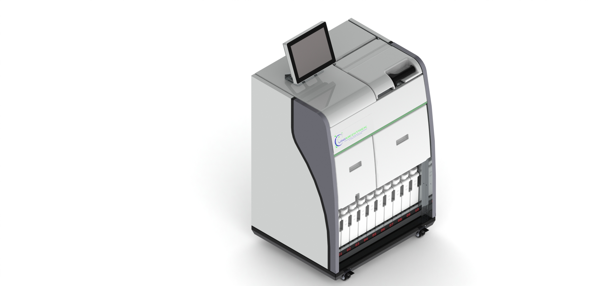
To avoid these pitfalls, using an Automatic Vacuum Tissue Processor can ensure consistent fixation by controlling the fixation time and pressure, enhancing tissue penetration. Following fixation, the Tissue Embedding Station can assist in the embedding process, ensuring that the tissue is adequately oriented for sectioning.
Conclusion and Practical Takeaway
Effective tissue fixation is a cornerstone of histopathology, and understanding the role of formalin, timing, and potential pitfalls is essential for achieving optimal results. By leveraging advanced equipment such as the Automatic Slide Staining Machine, Digital Tissue Floatation Bath, Slide Warming Table, and Cryostat Microtome, histopathology laboratories can enhance their workflow and improve diagnostic accuracy.
FAQs
What is the purpose of tissue fixation in histopathology?
Tissue fixation preserves cellular structure and composition for accurate microscopic examination.
How does timing affect tissue fixation?
Proper timing ensures that tissues are fixed adequately, preventing autolysis and preserving morphology.
Prepared by Unimeditrek Pvt. Ltd.. For product specifications and quotations, contact our team.