Home › Knowledge › Frozen Section Technique and Intra-Operative Workflow
Knowledge articleFrozen Section Technique and Intra-Operative Workflow
Understanding the essentials of frozen sectioning in histopathology
Introduction to Frozen Section Technique
The frozen section technique is a critical procedure in histopathology, allowing for rapid diagnosis during surgical operations. This method involves freezing tissue samples and slicing them into thin sections for microscopic examination, providing surgeons with immediate feedback on the presence of disease.
Intra-Operative Workflow
The workflow during a frozen section procedure is streamlined to ensure efficiency and accuracy. It typically involves the following steps:
- Sample Collection: Tissues are collected by the surgeon and sent to the pathology lab.
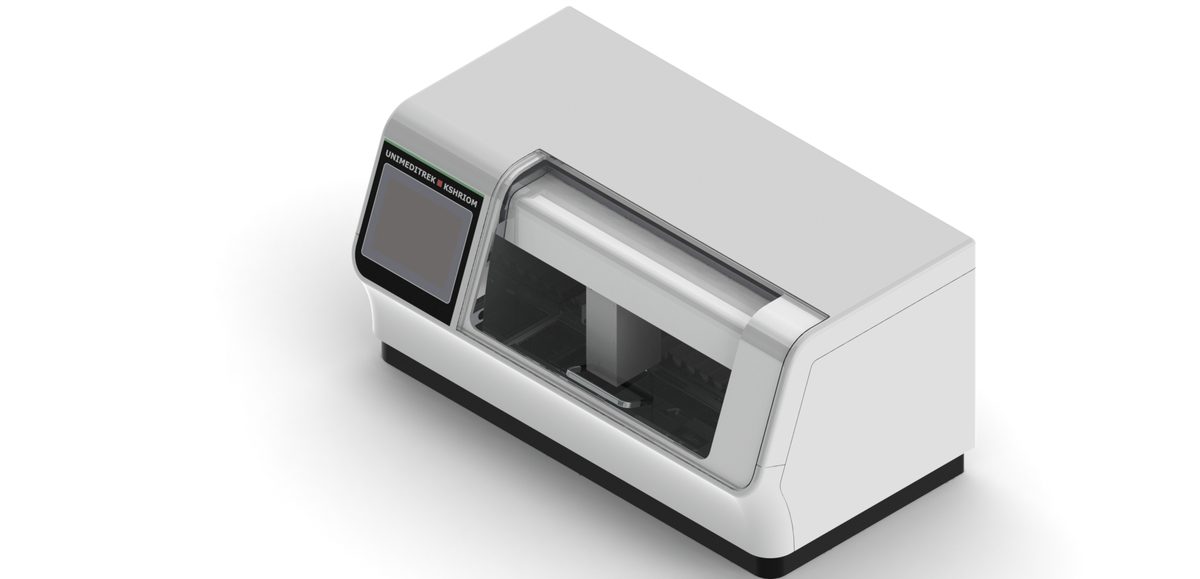
- Freezing: The Cryostat Microtome is used to rapidly freeze the tissue samples, preserving cellular architecture.
- Sectioning: The frozen tissue is sliced into thin sections using the Cryostat Microtome, ensuring optimal thickness for microscopic examination.
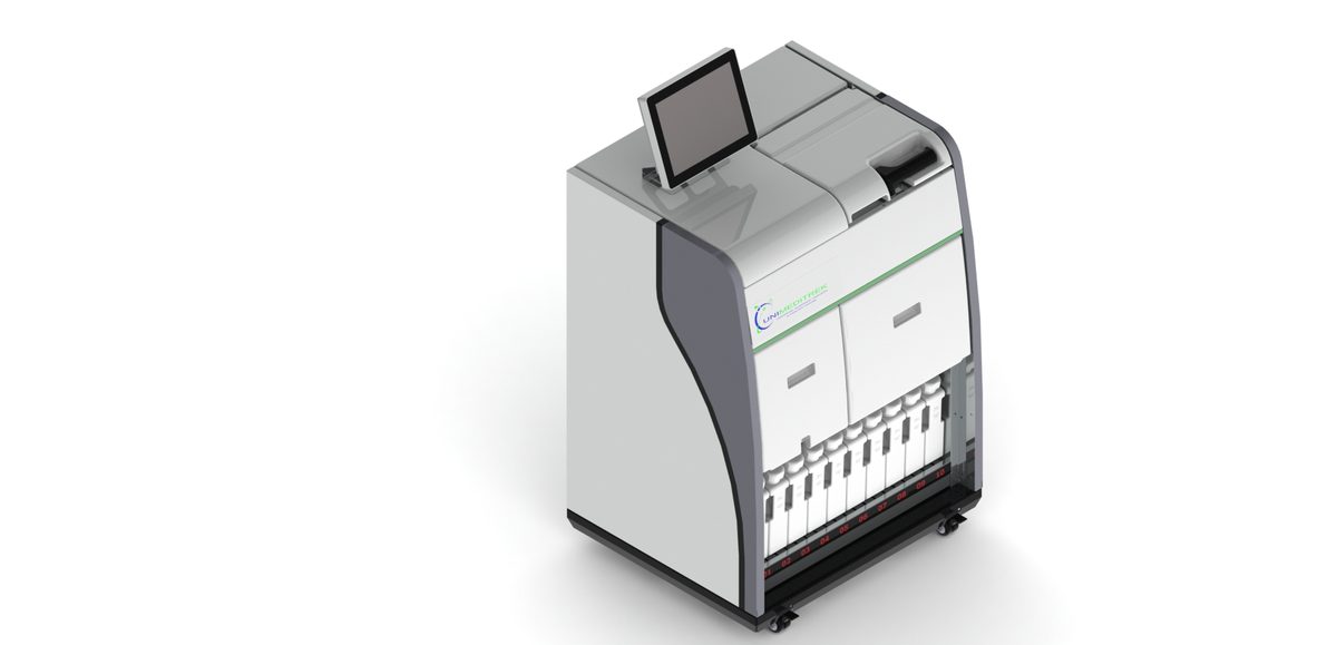
- Staining: The sections are then stained using the Automatic Slide Staining Machine for better visualization of cellular details.
- Microscopic Examination: Pathologists examine the stained slides, providing immediate diagnostic feedback.
Role of Equipment in Frozen Sectioning
Each piece of equipment plays a vital role in the success of the frozen section technique:
- Cryostat Microtome: This device is essential for producing high-quality frozen sections, allowing for precise cuts at varying thicknesses.
- Automatic Slide Staining Machine: It automates the staining process, saving time and ensuring consistency across samples.
- Tissue Embedding Station: While not directly involved in frozen sections, this equipment is crucial for embedding tissues in paraffin for subsequent analysis.
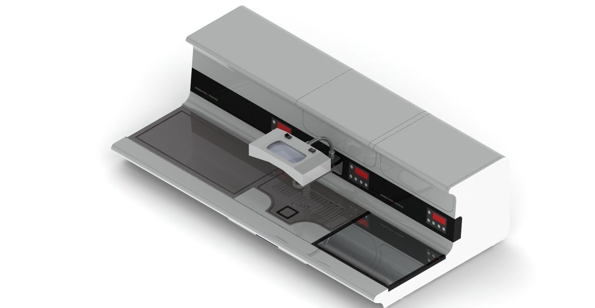
- Digital Tissue Floatation Bath: This bath aids in the proper flattening of tissue sections before staining, enhancing the quality of the slides.
- Slide Warming Table: This table ensures that slides are warmed to the right temperature, facilitating optimal staining and preventing damage to the tissue sections.
Challenges and Considerations
While the frozen section technique is invaluable, it comes with challenges such as potential ice crystal formation and the need for skilled personnel. Proper training and the use of high-quality equipment from Unimeditrek can mitigate these issues, ensuring accurate and timely diagnoses.
Practical Takeaway
Implementing the frozen section technique effectively requires a well-coordinated intra-operative workflow supported by reliable equipment. Investing in quality histopathology tools like the Cryostat Microtome and Automatic Slide Staining Machine from Unimeditrek can enhance diagnostic accuracy and improve patient outcomes.
FAQs
What is the frozen section technique?
The frozen section technique is a rapid diagnostic procedure that involves freezing tissue samples, slicing them, and examining them microscopically during surgery.
Why is the frozen section technique important?
It provides immediate diagnostic information to surgeons, allowing them to make critical decisions during operations.
Prepared and reviewed by the Unimeditrek technical team based on laboratory practice, installation and service experience. This document explains histopathology workflow, laboratory practice and equipment based on the questions we hear from hospitals, medical colleges and diagnostic laboratories — vendor-neutral where it explains the science and practical where it explains equipment.
Prepared by Unimeditrek Pvt. Ltd.. For product specifications and quotations, contact our team.