Home › Knowledge › Tissue Fixation in Histopathology: Formalin, Timing & Pitfalls
Knowledge articleTissue Fixation in Histopathology: Formalin, Timing & Pitfalls
A comprehensive guide to effective tissue fixation
Understanding Tissue Fixation
Tissue fixation is a crucial step in histopathology that preserves cellular structure and morphology, allowing for accurate diagnosis. The most commonly used fixative is formalin, a solution of formaldehyde in water. Proper fixation is essential to maintain the integrity of tissue samples, preventing degradation and ensuring reliable staining results.
The Role of Formalin in Tissue Fixation
Formalin works by cross-linking proteins, which stabilizes cellular structures. It penetrates tissues quickly, but its effectiveness can be influenced by several factors including concentration, temperature, and pH. Typically, a 10% neutral buffered formalin (NBF) is used, which balances fixation efficiency and tissue preservation.
Timing: The Key to Effective Fixation
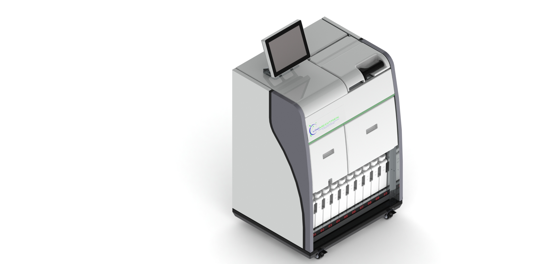
The duration of fixation is critical. Under-fixation can lead to poor staining, while over-fixation can cause tissue hardening and loss of antigenicity. Ideal fixation time varies depending on tissue type and thickness, but generally, small biopsies require 6-8 hours, while larger specimens may need 24 hours or more. Employing an Automatic Vacuum Tissue Processor can help standardize fixation times and improve consistency across samples.
Common Pitfalls in Tissue Fixation
Several pitfalls can compromise tissue fixation:
- Inadequate penetration: Thick tissues may not fix uniformly, leading to artifacts.
- Delayed fixation: Leaving tissues in formalin for too long can alter morphology.
- Improper temperature: Fixatives should be kept at room temperature to ensure optimal performance.
To avoid these issues, it’s important to monitor fixation conditions closely and utilize reliable equipment. For instance, the Tissue Embedding Station can assist in the embedding process post-fixation, ensuring that samples are processed efficiently.
Conclusion and Practical Takeaway
Effective tissue fixation is vital for accurate histopathological diagnosis. By understanding the role of formalin, adhering to optimal timing, and avoiding common pitfalls, professionals can enhance the quality of their samples. Incorporating advanced tools like the Automatic Slide Staining Machine and Cryostat Microtome can further streamline the histopathology workflow, leading to better outcomes.
FAQs
What is the ideal fixation time for different tissue types?
Fixation time varies; small biopsies typically need 6-8 hours, while larger specimens may require 24 hours or more.
How can common pitfalls in tissue fixation be avoided?
Monitor fixation conditions, ensure proper penetration, and use reliable equipment like the Automatic Vacuum Tissue Processor.
This document has been prepared and reviewed by the Unimeditrek technical team based on histopathology workflow, laboratory practice, installation and service experience, and questions we hear from hospitals, medical colleges and diagnostic laboratories. It is vendor-neutral where it explains the science and practical where it explains equipment.
Prepared by Unimeditrek Pvt. Ltd.. For product specifications and quotations, contact our team.