Home › Knowledge › H&E Staining Theory and Standardization Guide
Knowledge articleH&E Staining Theory and Standardization Guide
Mastering Hematoxylin and Eosin Staining
Understanding H&E Staining
Hematoxylin and Eosin (H&E) staining is a fundamental technique in histopathology, widely used to visualize tissue morphology. Hematoxylin stains cell nuclei blue, while Eosin imparts a pink hue to the cytoplasm and extracellular matrix. This dual staining provides a comprehensive view of tissue architecture, aiding in the diagnosis of various diseases.
The Importance of Standardization
Standardizing the H&E staining process is crucial for ensuring reproducibility and accuracy in histopathological assessments. Variability in staining can lead to misinterpretation of results, affecting diagnosis and treatment decisions. Standardization involves controlling various factors, including reagent quality, staining time, and temperature.
Key Steps for Standardizing H&E Staining
- Reagent Quality: Use high-quality hematoxylin and eosin reagents to ensure consistent staining results. Regularly check expiration dates and storage conditions.
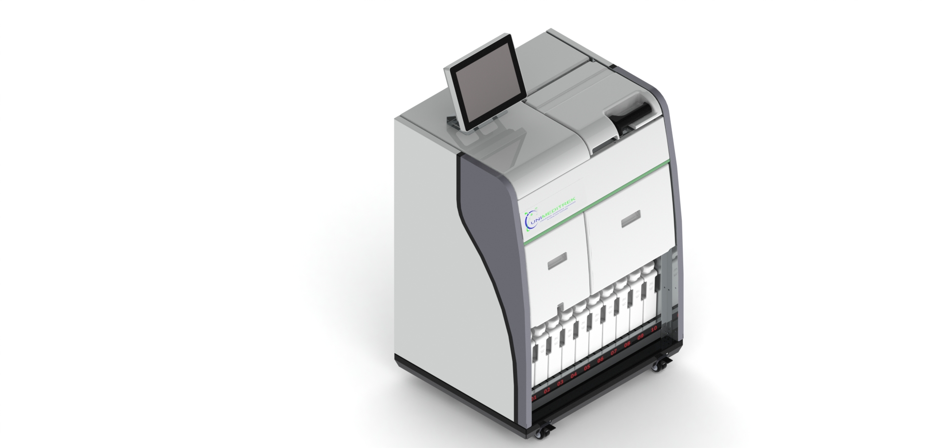
- Consistent Timing: Adhere to standardized staining times for each step. Utilize an Automatic Slide Staining Machine to maintain precise timing.
- Temperature Control: Maintain a consistent temperature during staining. A Slide Warming Table can help achieve optimal conditions for drying slides.
- Embedding Techniques: Proper embedding of tissues is critical. Use a Tissue Embedding Station to ensure consistent infiltration and embedding of tissues.
- Floating Sections: Utilize a Digital Tissue Floatation Bath for optimal sectioning and to prevent tissue damage during transfer to slides.
Utilizing Advanced Equipment
Incorporating advanced equipment can greatly enhance the standardization of H&E staining. The Automatic Vacuum Tissue Processor ensures uniform processing of tissue samples, reducing variability. Additionally, a Cryostat Microtome offers precise sectioning of frozen tissues, which is essential for certain diagnostic procedures.
Conclusion and Practical Takeaway
Standardizing H&E staining is vital for achieving reliable histopathological results. By focusing on reagent quality, timing, temperature control, and utilizing advanced equipment, laboratories can enhance the accuracy of their histological assessments. Investing in tools like the Automatic Slide Staining Machine and the Tissue Embedding Station will streamline the process and improve overall outcomes.
FAQs
What is H&E staining used for?
H&E staining is used to visualize tissue morphology and diagnose diseases by highlighting cell nuclei and cytoplasm.
How can I improve the consistency of my H&E staining?
Improve consistency by standardizing reagent quality, timing, temperature, and using advanced equipment like automatic slide staining machines.
This document has been prepared and reviewed by the Unimeditrek technical team based on histopathology workflow, laboratory practice, installation and service experience, and questions we hear from hospitals, medical colleges and diagnostic laboratories. It is vendor-neutral where it explains the science and practical where it explains equipment.
Prepared by Unimeditrek Pvt. Ltd.. For product specifications and quotations, contact our team.