Home › Knowledge › Decalcification Methods for Bone and Calcified Specimens
Knowledge articleDecalcification Methods for Bone and Calcified Specimens
Understanding Decalcification Techniques in Histopathology
Introduction to Decalcification
Decalcification is a critical step in the preparation of bone and calcified specimens for histopathological examination. This process involves the removal of calcium salts to facilitate the slicing of tissues into thin sections, enabling accurate microscopic analysis. Without effective decalcification, the integrity of the tissue can be compromised, leading to diagnostic errors.
Common Decalcification Methods
There are several methods employed for decalcification, each with its advantages and limitations:
- Chemical Decalcification: This is the most common method, utilizing acids such as hydrochloric acid or nitric acid. The tissue is submerged in the decalcifying solution until the calcium is adequately removed.
- Electrolytic Decalcification: This method uses an electric current to accelerate the decalcification process. It is faster than chemical methods but requires specialized equipment.
- Ultrasonic Decalcification: This technique employs ultrasound waves to enhance the penetration of the decalcifying agent, speeding up the process.
Factors Influencing Decalcification
Several factors can affect the efficiency of decalcification:
- Type of Acid: The choice of acid can influence the speed and effectiveness of calcium removal.
- Temperature: Warmer temperatures can enhance the reaction rate, but excessive heat can damage the tissue.
- Time: Overexposure to decalcifying agents can lead to tissue degradation, while insufficient time may leave calcium deposits.
Integrating Equipment for Effective Decalcification
Utilizing advanced histopathology equipment can significantly enhance the decalcification process:
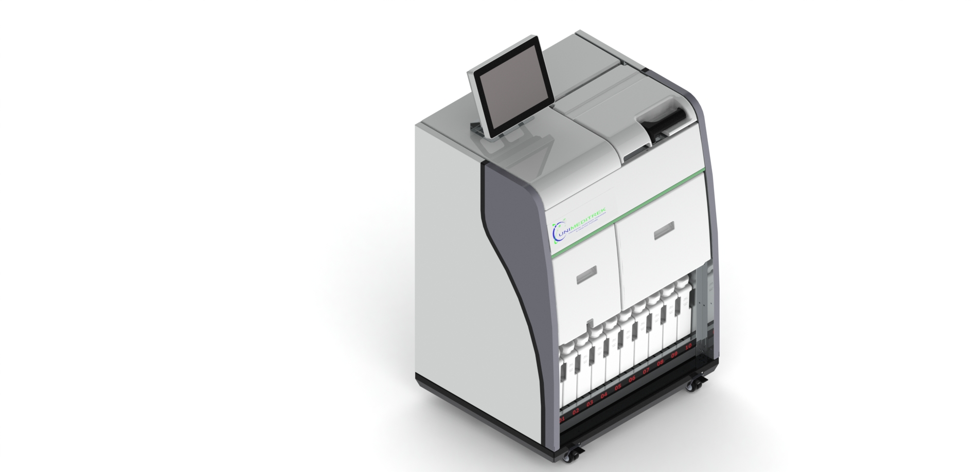
- Automatic Vacuum Tissue Processor: This equipment can automate the decalcification process, ensuring consistent exposure to the decalcifying agent and reducing manual errors.
- Tissue Embedding Station: After decalcification, proper embedding is essential for preserving tissue morphology. An embedding station ensures optimal orientation and support for the specimen.
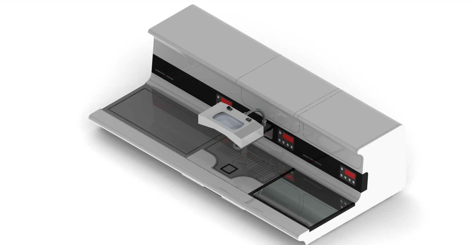
- Automatic Slide Staining Machine: Once decalcified, staining is crucial for visualizing tissue structures. An automatic slide staining machine provides uniform staining, enhancing diagnostic accuracy.
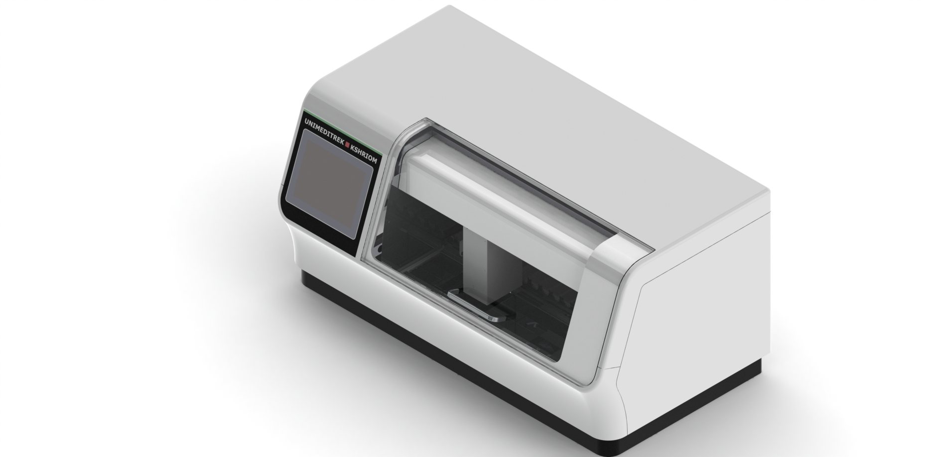
- Cryostat Microtome: For cutting thin sections of decalcified tissues, a cryostat microtome allows for precise slicing, preserving the integrity of the specimens.
Conclusion and Practical Takeaway
Decalcification is an essential process in histopathology, particularly for bone and calcified specimens. By understanding the various methods and employing modern equipment like Unimeditrek's Automatic Vacuum Tissue Processor and Cryostat Microtome, laboratories can achieve high-quality tissue sections, leading to better diagnostic outcomes.
FAQs
What is decalcification in histopathology?
Decalcification is the process of removing calcium salts from bone and calcified tissues to prepare them for microscopic examination.
What equipment can assist in the decalcification process?
Equipment such as the Automatic Vacuum Tissue Processor and Cryostat Microtome can enhance the efficiency and quality of the decalcification process.
This document has been prepared and reviewed by the Unimeditrek technical team based on histopathology workflow, laboratory practice, installation and service experience, and questions we hear from hospitals, medical colleges and diagnostic laboratories. It is vendor-neutral where it explains the science and practical where it explains equipment.
Prepared by Unimeditrek Pvt. Ltd.. For product specifications and quotations, contact our team.