Home › Knowledge › Automatic Microtome in Daily Histopathology
Use caseAutomatic Microtome in Daily Histopathology
Streamlining Tissue Sectioning for Accurate Diagnosis
Introduction to the Automatic Microtome
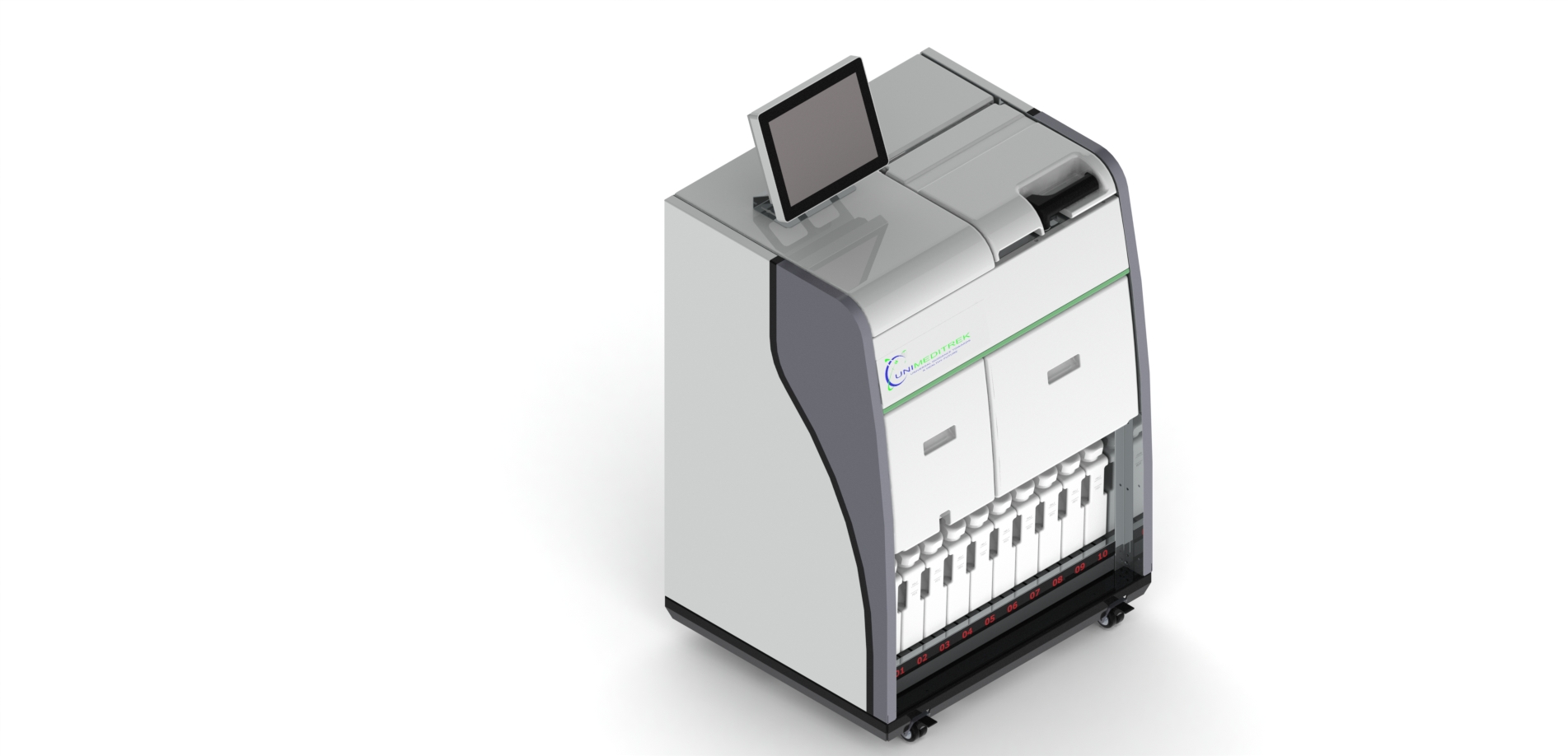
The Automatic Microtome is a vital instrument in histopathology, used for slicing thin sections of tissue for microscopic examination. In a typical histopathology lab, where precision and speed are crucial, the Automatic Microtome stands out as an essential tool that enhances the workflow and accuracy of tissue processing.
Daily Operations in a Histopathology Lab
In a busy histopathology lab, the day begins with the preparation of tissue samples that have been fixed and embedded in paraffin. The Automatic Microtome is set up to ensure that the slicing process is efficient and consistent. Technicians load the paraffin-embedded tissue blocks into the microtome, adjusting the settings for thickness based on the specific requirements of the analysis. Typically, sections are cut at a thickness of 3 to 5 micrometers, which allows for optimal visualization under the microscope.
Benefits of Using an Automatic Microtome
The Automatic Microtome offers several advantages over manual microtomes. Firstly, it significantly reduces the time taken to prepare tissue sections, allowing pathologists to receive samples more quickly. This speed is particularly beneficial in high-volume labs where timely diagnosis can impact patient care. Secondly, the precision of the Automatic Microtome ensures uniformity in section thickness, which is critical for accurate staining and subsequent analysis. Lastly, the automation reduces the physical strain on technicians, as they do not need to manually operate the device for each cut.
Quality Control and Maintenance
To maintain the quality of the tissue sections produced, routine calibration and maintenance of the Automatic Microtome are essential. Technicians are trained to perform daily checks, including blade sharpness and alignment, to ensure optimal performance. Regular cleaning of the microtome also prevents contamination and ensures that the sections remain intact during the cutting process. By adhering to these practices, labs can maximize the longevity of their equipment and the quality of their diagnostic results.
Conclusion and Practical Takeaway
In conclusion, the Automatic Microtome is an indispensable tool in histopathology labs across India, streamlining the process of tissue sectioning and improving diagnostic efficiency. By embracing this technology, labs can enhance their workflow and provide timely, accurate results that are crucial for patient management. For technicians and pathologists alike, understanding the operation and maintenance of the Automatic Microtome is key to optimizing its benefits in daily practice.
FAQs
What is an Automatic Microtome?
An Automatic Microtome is a device used to cut thin sections of tissue for microscopic examination, enhancing precision and efficiency in histopathology.
How does the Automatic Microtome improve lab efficiency?
It reduces the time required for sectioning, ensures uniformity in thickness, and minimizes physical strain on technicians, thereby streamlining the workflow.
This document has been prepared and reviewed by the Unimeditrek technical team based on histopathology workflow, laboratory practice, installation and service experience, and questions we hear from hospitals, medical colleges and diagnostic laboratories. It is vendor-neutral where it explains the science and practical where it explains equipment.
Prepared by Unimeditrek Pvt. Ltd.. For product specifications and quotations, contact our team.