Home › Knowledge › Artifact Identification in Histology Sections and Prevention
Knowledge articleArtifact Identification in Histology Sections and Prevention
Understanding and Mitigating Histological Artifacts
Understanding Histological Artifacts
In histopathology, artifacts are distortions or alterations in tissue sections that can lead to misinterpretation of results. These artifacts can arise from various stages of tissue processing, embedding, sectioning, and staining. Common types of artifacts include shrinkage, folding, and staining irregularities, each of which can obscure true tissue morphology.
Common Types of Artifacts
- Shrinkage: Often caused by dehydration during processing, leading to gaps between cells.
- Folding: Occurs during sectioning, where the tissue folds over itself, complicating the examination.
- Staining Artifacts: These arise from improper slide staining, such as uneven dye distribution or precipitates.
- Air Bubbles: These can form during embedding or staining, creating voids that mimic pathological changes.
Preventing Artifacts in Histology Sections
Preventing artifacts requires meticulous attention to detail at every stage of tissue preparation. Here are some effective strategies:
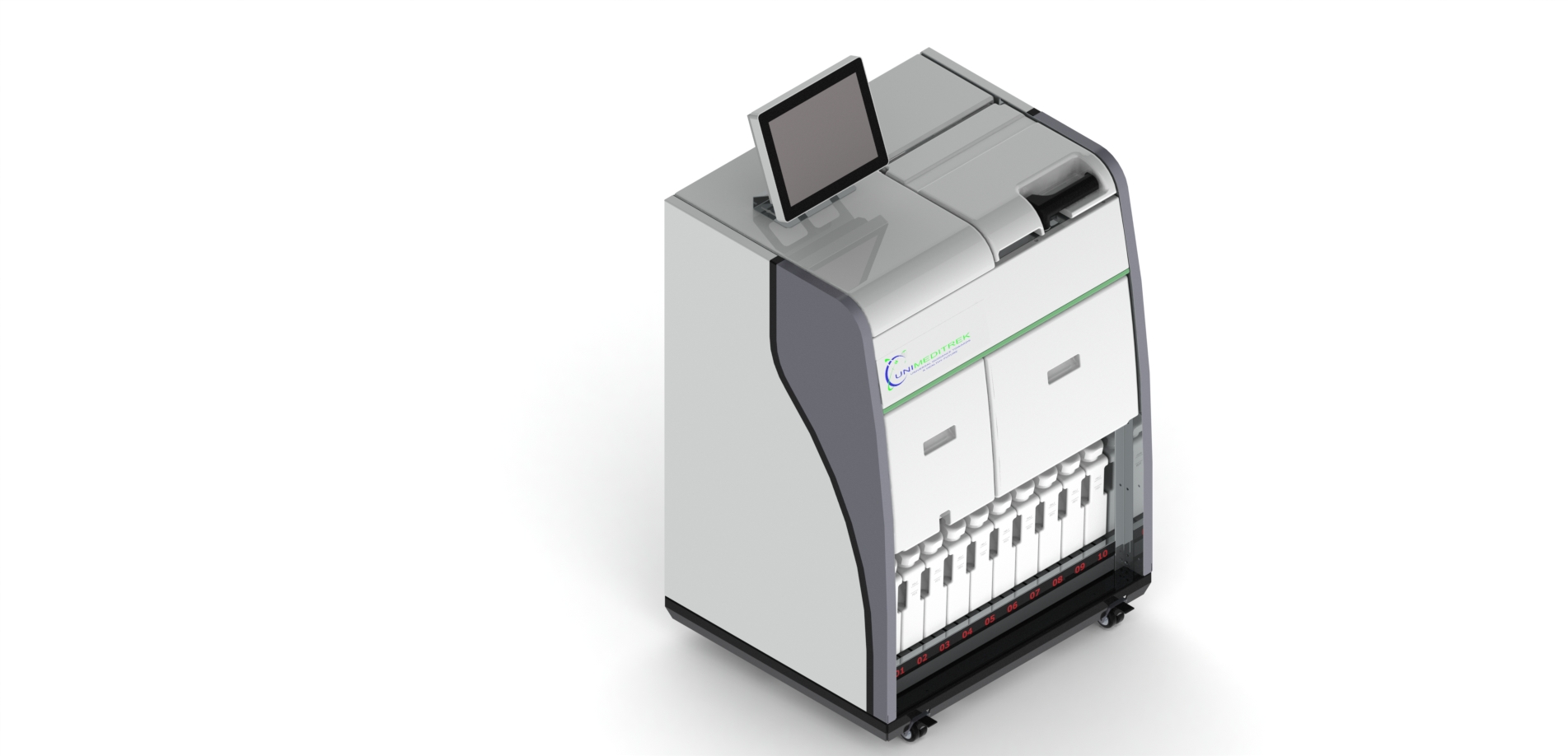
- Use of Automatic Vacuum Tissue Processor: This equipment ensures consistent dehydration and infiltration, reducing shrinkage artifacts.
- Tissue Embedding Station: Proper embedding techniques help maintain tissue architecture, minimizing folding and distortion.
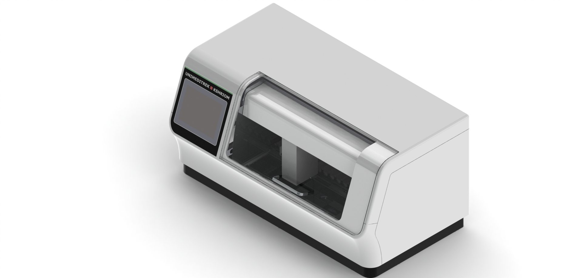
- Automatic Slide Staining Machine: Automated staining provides uniform application of reagents, reducing staining artifacts.
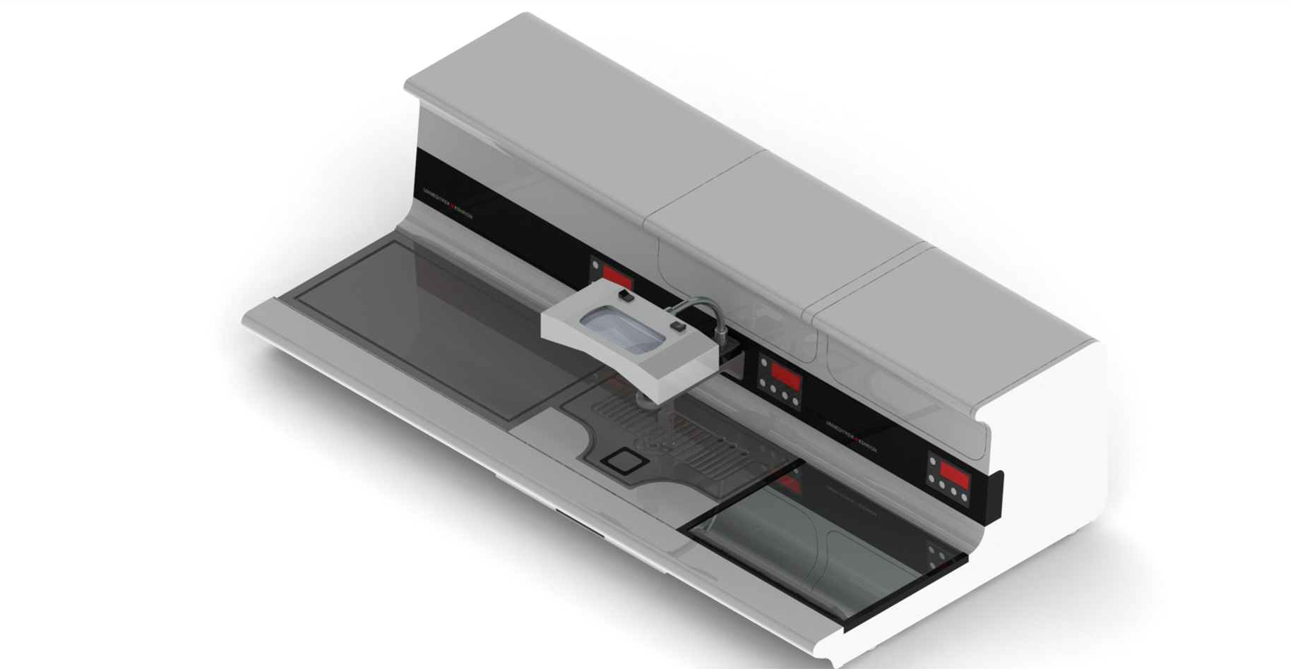
- Digital Tissue Floatation Bath: This device aids in flattening sections, preventing folds and ensuring optimal morphology.
- Slide Warming Table: A controlled environment for drying slides can help prevent air bubbles and ensure even staining.
- Cryostat Microtome: For frozen sections, this equipment allows for precise cutting, reducing artifacts associated with paraffin embedding.
Best Practices for Artifact Identification
To effectively identify artifacts, histopathologists should develop a keen eye for detail. Regular training and quality control measures can enhance the ability to distinguish between true pathological changes and artifacts. Additionally, maintaining a clean workspace and using high-quality reagents can significantly reduce the occurrence of artifacts.
Practical Takeaway
Identifying and preventing artifacts in histology is crucial for accurate diagnosis and research outcomes. By utilizing advanced equipment like the Automatic Vacuum Tissue Processor and the Automatic Slide Staining Machine, histopathologists can significantly reduce the risk of artifacts and improve the reliability of their findings.
FAQs
What are common artifacts found in histology sections?
Common artifacts include shrinkage, folding, staining irregularities, and air bubbles.
How can I prevent artifacts in my histology lab?
Utilize automated equipment like vacuum tissue processors and slide staining machines to ensure consistent processing and staining.
This document has been prepared and reviewed by the Unimeditrek technical team based on histopathology workflow, laboratory practice, installation and service experience, and questions we hear from hospitals, medical colleges and diagnostic laboratories. It is vendor-neutral where it explains the science and practical where it explains equipment.
Prepared by Unimeditrek Pvt. Ltd.. For product specifications and quotations, contact our team.